100. Plasma Proteins: Classification, Functions, and Electrophoresis
Blood is thicker than water, but it is the invisible dissolved molecules that truly sustain life. When looking at a vial of blood, one might only consider red blood cells, but the clear liquid left behind holds a complex, highly regulated biochemical universe. This slide deck explores the fundamental nature, structure, and function of this vital system. Designed for medical and biochemistry students, it demystifies complex physiological processes, making clinical analysis and molecular interactions accessible and engaging.
Slide 1: Introduction to the Biochemistry of Plasma Proteins
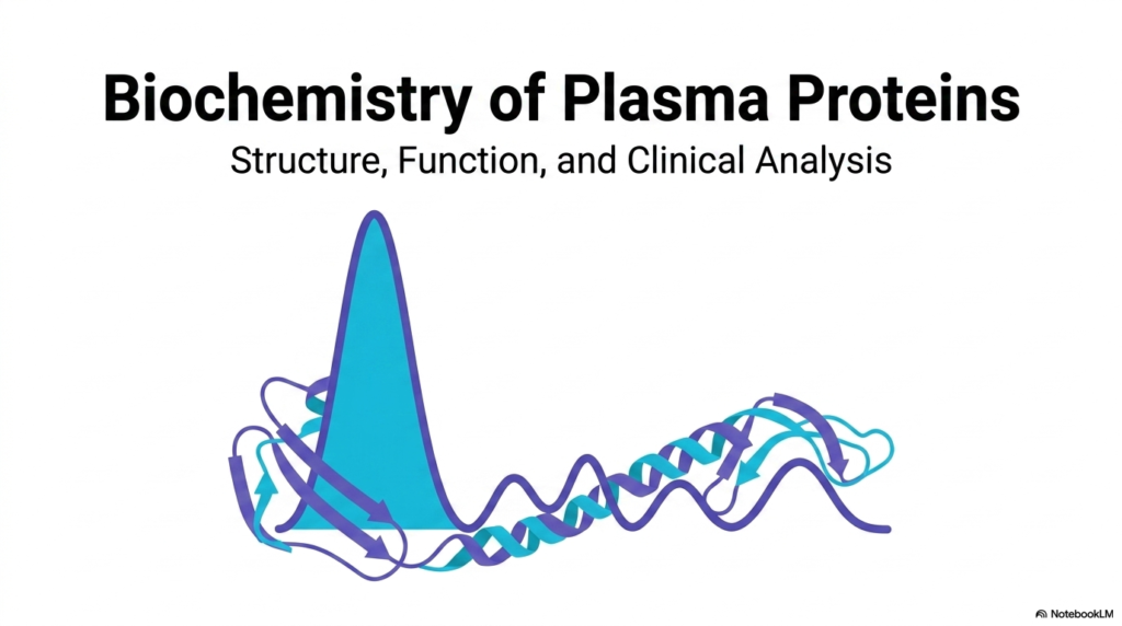
The first slide introduces the expansive and intricate world of plasma proteins, establishing a strong foundation for the biochemical analysis that follows. It immediately highlights the vital intersection of molecular structure, biological function, and clinical diagnostic methods. Students observing this title slide are presented with an elegant visual representation of a classic electrophoresis curve overlapping a stylized alpha-helical protein ribbon. This striking imagery perfectly encapsulates the dual nature of the lessons ahead. By studying plasma proteins, future medical professionals learn how invisible, microscopic molecular structures directly translate into measurable, macroscopic clinical data.
A fundamental theme introduced here is that plasma proteins do not exist as static, inert building blocks. Instead, they constitute a highly dynamic, tightly regulated physiological system requiring continuous synthesis, deployment, and eventual degradation. The stylized protein structure on the slide hints at the complex polypeptide folding, secondary structures, and tertiary conformations that completely define their biological functionality. From maintaining vascular osmotic pressure to defending against microscopic pathogens, these specific macromolecules are absolutely indispensable for human survival. Their structural integrity dictates their ability to perform life-sustaining biochemical tasks.
The prominent graphical curve superimposed over the protein structure represents the clinical reality of analyzing these components in a laboratory setting. Medical practitioners constantly rely on understanding the normal distributions of plasma proteins to detect systemic biochemical imbalances or hidden underlying pathologies. The large initial peak, universally recognized as albumin, acts as a visual anchor point for interpreting the entire electrophoretic spectrum. This introductory slide establishes the clear expectation that upcoming concepts will continually bridge the gap between abstract molecular theory and practical clinical application.
Ultimately, the study of plasma proteins provides a profound window into the overall health and metabolic stability of the human body. Mastering the structural properties and clinical distributions of these molecules is the very first step toward utilizing the sophisticated diagnostic tools central to modern medical practice. The subsequent slides will dissect this complex system, revealing exactly how individual protein fractions contribute to the broader physiological harmony required to sustain life.
Slide 2: The Circulating Proteome and Plasma Proteins
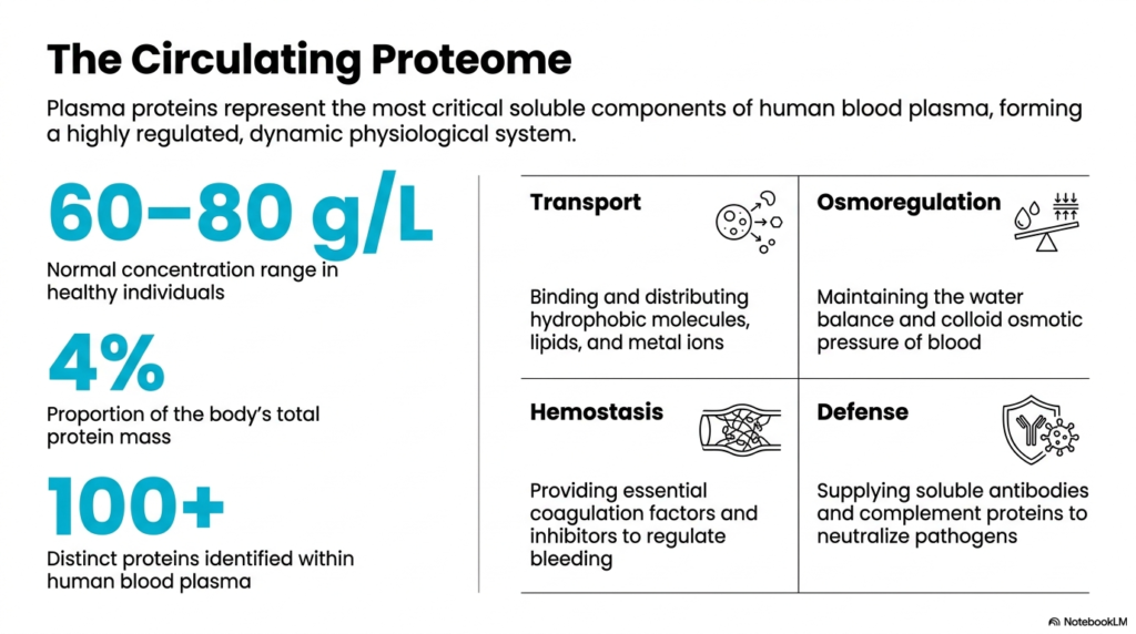
The second slide transitions into the quantitative reality of the circulating proteome, defining the exact parameters that govern plasma proteins in healthy individuals. The human body tightly regulates these soluble components, maintaining a strict concentration range between sixty and eighty grams per liter of blood. Although they represent only four percent of the body’s total protein mass, their physiological impact is disproportionate to their weight. Over one hundred distinct molecules have been identified within this fluid matrix, each performing highly specialized tasks necessary for metabolic stability.
To comprehend the true value of plasma proteins, one must examine their four primary physiological roles detailed on this slide. The first critical function is transport. These macromolecules act as biological ferries, safely binding and distributing hydrophobic molecules, essential lipids, and reactive metal ions through the aqueous environment of the bloodstream. Without this transport mechanism, vital insoluble nutrients would simply precipitate out of the blood, and toxic ions would cause widespread cellular damage.
The second and third functions highlight the structural and protective roles of plasma proteins. Osmoregulation relies heavily on these molecules to maintain water balance and blood colloid osmotic pressure, preventing fluid from indiscriminately leaking into surrounding tissues. Simultaneously, hemostasis depends on a delicate balance of coagulation factors and biochemical inhibitors provided by this protein matrix. These factors stand ready to initiate complex enzymatic cascades the moment vascular injury occurs, effectively preventing fatal hemorrhage while also stopping abnormal clotting.
Finally, the defense mechanism showcases the remarkable immune capabilities of plasma proteins. Soluble antibodies and complement system components constantly circulate, seeking out and neutralizing foreign pathogens before they can establish an infection. By breaking down these four pillars—transport, osmoregulation, hemostasis, and defense—students gain a comprehensive understanding of why maintaining normal protein concentrations is paramount. Any deviation from these established baselines immediately signals clinical distress, emphasizing the critical diagnostic value of the circulating proteome.
Slide 3: The Principal Biochemical Division of Plasma Proteins
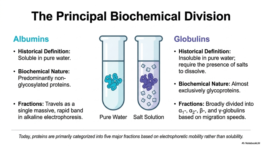
Slide three outlines the fundamental biochemical classification system used to categorize plasma proteins, distinguishing between the two primary groups: albumins and globulins. Historically, this division was based entirely on basic solubility principles. Albumins were identified by their unique ability to remain completely soluble in pure water. In stark contrast, globulins were defined by their insolubility in pure water, requiring the specific presence of salt solutions to properly dissolve. While modern science relies on advanced techniques, these historical definitions still provide valuable insight into their distinct physical properties.
The true biochemical nature of these plasma proteins reveals far more than just solubility differences. Albumins stand out because they are predominantly non-glycosylated proteins, meaning they lack attached carbohydrate chains. This unique structural simplicity allows the albumin fraction to travel as a single, massive, and highly rapid band during alkaline carrier electrophoresis. Because of its compact and highly charged nature, it outpaces all other molecules when subjected to an electrical field, making it the most easily identifiable component in any clinical serum analysis.
Conversely, the globulin group represents a highly diverse and complex family of plasma proteins that are almost exclusively glycoproteins. The heavy glycosylation—the attachment of complex sugar structures to the protein backbone—significantly alters their mass, charge, and overall behavior in the bloodstream. Because of this structural diversity, globulins do not migrate as a unified group. Instead, they broadly separate into four distinct fractions based on their individual migration speeds: alpha-1, alpha-2, beta, and gamma globulins.
Today, modern clinical biochemistry prioritizes categorization based on these specific electrophoretic mobilities rather than archaic solubility tests. By observing how plasma proteins separate under an electrical current, biochemists can accurately isolate and quantify each functional group. Understanding this principal division between the streamlined albumins and the heavily glycosylated globulins is absolutely essential for interpreting complex electrophoretic patterns. This foundational knowledge allows medical students to accurately connect molecular structures to their corresponding diagnostic bands.
Slide 4: Serum Albumin as the Predominant of Plasma Proteins
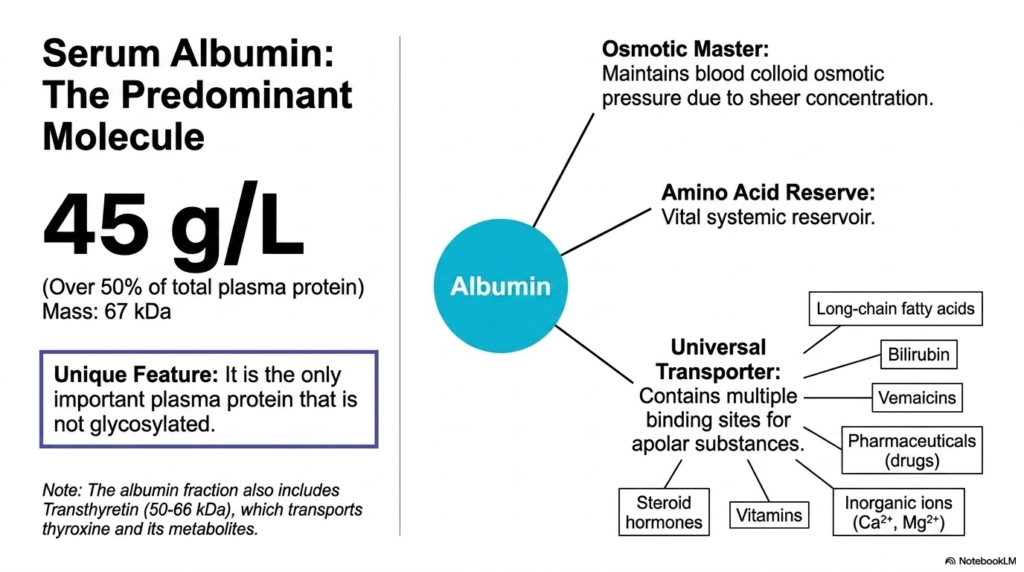
Slide four focuses exclusively on serum albumin, unquestionably the most abundant and vital component among all plasma proteins. Presenting at a massive concentration of forty-five grams per liter, albumin constitutes more than fifty percent of the total protein mass in human plasma. Despite its relatively modest molecular weight of sixty-seven kilodaltons, its sheer volume grants it immense physiological power. A uniquely defining feature of this specific molecule is its complete lack of glycosylation, distinguishing it from nearly every other major protein circulating in the human bloodstream.
The primary physiological role of this singular molecule is serving as the absolute master of osmoregulation. Because it exists in such overwhelming concentrations, albumin is almost entirely responsible for maintaining the blood colloid osmotic pressure. This oncotic pressure creates a vital biochemical pull that keeps water safely contained within the vascular system. If the concentration of these specific plasma proteins drops due to liver failure or malnutrition, fluid rapidly escapes into surrounding tissues, resulting in severe, visible clinical edema.
Beyond osmotic control, albumin functions as the body’s premier universal transporter. Its unique three-dimensional structure features multiple high-capacity binding sites specifically designed to capture and carry a vast array of apolar substances. This incredible molecule safely transports long-chain fatty acids, potentially toxic bilirubin, various steroid hormones, and essential inorganic ions like calcium and magnesium. Furthermore, modern medicine relies heavily on albumin to distribute pharmaceutical drugs efficiently throughout the body, making it a critical factor in human pharmacology.
Additionally, the albumin fraction serves as a vital systemic amino acid reserve, providing emergency nutritional building blocks during times of severe metabolic starvation. It is also important to note that the broader albumin electrophoretic fraction technically includes transthyretin, a smaller specialized molecule responsible for transporting thyroxine and its metabolites. Mastering the multifaceted roles of these predominant plasma proteins is absolutely crucial for any medical student, as albumin influences nearly every aspect of human biochemistry and fluid dynamics.
Slide 5: The Globulin Workforce Within Plasma Proteins
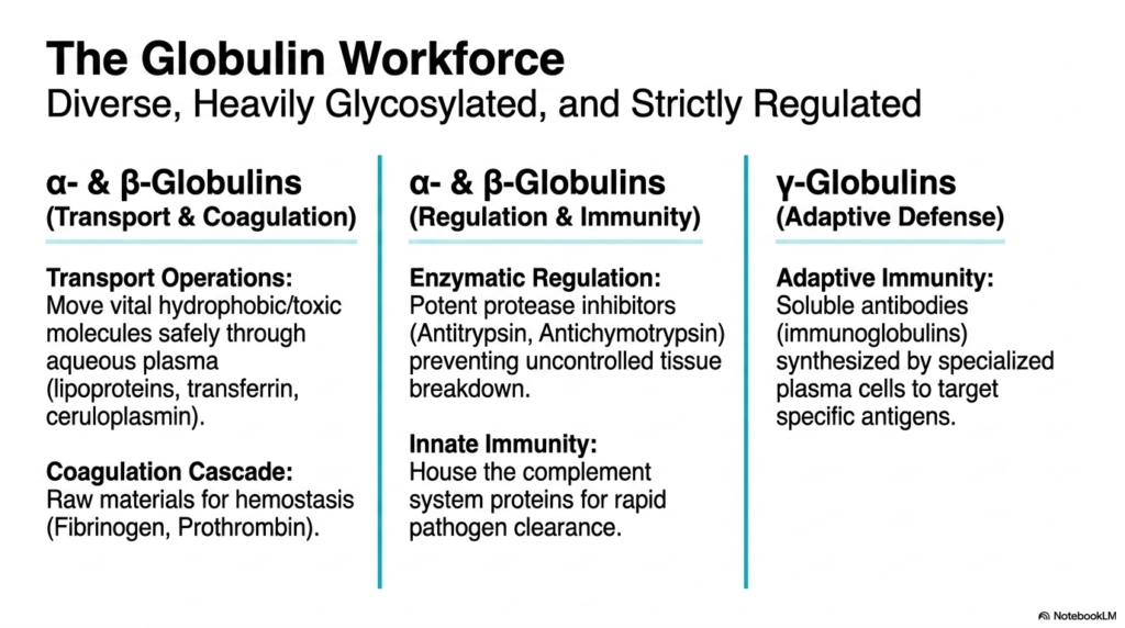
Slide five shifts focus to the diverse, heavily glycosylated, and strictly regulated molecules known as globulins, which constitute the remaining fractions of plasma proteins. Unlike the singular dominance of albumin, the globulin family is divided into highly specialized subgroups, each managing distinct physiological functions. The alpha and beta globulins primarily serve dual roles in complex transport operations and the critical coagulation cascade. These molecules safely move vital hydrophobic compounds, toxic molecules, and trace metals through the hostile aqueous environment of human blood plasma.
A critical subset of these specific plasma proteins functions as regulatory enzymes, primarily acting as potent protease inhibitors. Molecules like antitrypsin and antichymotrypsin are absolutely essential for preventing the uncontrolled breakdown of healthy bodily tissues. Without these regulating globulins constantly circulating in the blood, naturally occurring digestive enzymes would aggressively attack internal organs, leading to catastrophic systemic damage. Furthermore, the alpha and beta fractions house the crucial complement system, providing rapid innate immunity to instantly clear foreign pathogens upon detection.
Simultaneously, the alpha and beta globulins act as the essential raw materials required for hemostasis. Key precursor molecules, including fibrinogen and prothrombin, circulate inertly until triggered by vascular injury. Once activated, they form the structural foundation of blood clots. This demonstrates how carefully the human body regulates its internal chemistry, ensuring that these plasma proteins remain inactive during normal circulation but instantly mobilize to prevent fatal hemorrhage during physical trauma.
Finally, the slide introduces the gamma globulins, which are strictly dedicated to adaptive defense. Unlike other fractions, these plasma proteins consist entirely of soluble antibodies, or immunoglobulins. They are uniquely synthesized by specialized immune cells rather than the liver and are designed to target and remember exact antigens. This division of labor—where alpha and beta handle transport and regulation, while gamma manages advanced immunity—illustrates the incredible biochemical sophistication of the globulin workforce in maintaining human health.
Slide 6: Targeted Transport Systems Using Plasma Proteins
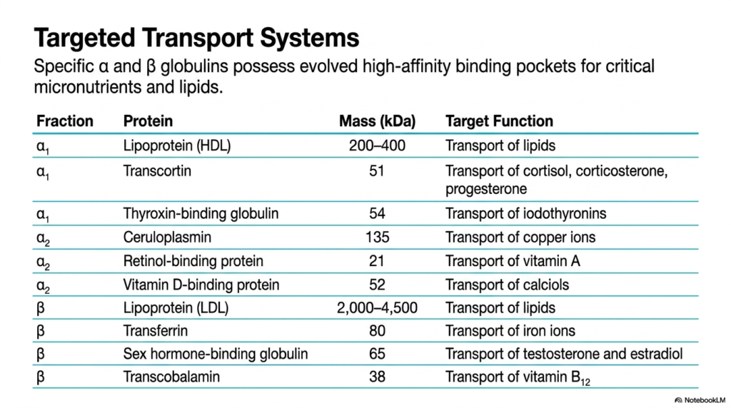
Slide six provides a highly detailed breakdown of how specific alpha and beta globulins function as evolved, targeted transport systems within the realm of plasma proteins. While albumin acts as a general cargo carrier, these specific globulins possess uniquely evolved, high-affinity binding pockets designed for very exact micronutrients and lipids. This specialization ensures that highly reactive, delicate, or completely insoluble molecules can safely travel through the circulatory system without degrading or causing dangerous peripheral toxicity to the surrounding vascular tissues.
Within the alpha-1 fraction, specialized plasma proteins handle distinct and vital endocrine and metabolic cargo. High-density lipoproteins, carrying a massive weight of up to four hundred kilodaltons, manage the crucial transport of cellular lipids. Transcortin is specifically engineered to safely transport powerful steroid hormones like cortisol and progesterone. Similarly, thyroxin-binding globulin strictly manages the distribution of iodothyronines, ensuring thyroid hormones reach their target receptors completely intact. This precise molecular targeting prevents chaotic hormonal signaling throughout the human body.
The alpha-2 fraction contains heavy-duty transporters essential for trace metal and vitamin regulation. Ceruloplasmin, a massive molecule, is solely responsible for isolating and transporting reactive copper ions, preventing them from generating dangerous free radicals in the blood. Retinol-binding protein and vitamin D-binding protein carefully carry crucial fat-soluble vitamins to tissues that desperately require them for cellular maintenance and bone health. Each of these specific plasma proteins demonstrates an incredible level of evolutionary design targeted at maintaining precise systemic homeostasis.
Moving to the beta fraction, students encounter massive structural carriers like low-density lipoproteins, which can reach an astonishing four million five hundred thousand daltons in mass. Transferrin strictly regulates iron ion transport, while sex hormone-binding globulin carefully distributes testosterone and estradiol. Transcobalamin specifically ensures the delivery of vitamin B12. Understanding these targeted plasma proteins is vital for medical students, as a deficiency in any single carrier leads to immediate, identifiable, and often severe clinical metabolic diseases.
Slide 7: Hemostasis, Inhibition, and Reactivity of Plasma Proteins
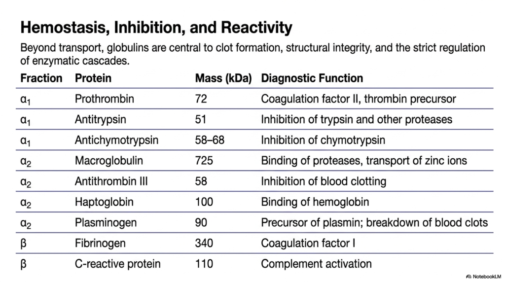
Slide seven explores the crucial non-transport functions of globulins, focusing heavily on how these specific plasma proteins manage clot formation, structural integrity, and the strict regulation of enzymatic cascades. Beyond simply carrying molecules, the alpha and beta globulins are absolutely central to survival during physical trauma and inflammation. They achieve this through a highly complex, interconnected web of precursor molecules and aggressive biochemical inhibitors that constantly monitor and manipulate the physical state of human blood.
The alpha-1 fraction houses essential regulatory plasma proteins like antitrypsin and antichymotrypsin. These fifty-to-sixty kilodalton molecules act as potent inhibitors of trypsin and chymotrypsin, effectively neutralizing rogue proteases that could inappropriately digest healthy vascular tissue. Similarly, the alpha-2 fraction contains massive regulatory molecules like macroglobulin, which acts as a molecular trap to bind proteases and transport zinc ions, alongside antithrombin III, which is absolutely vital for inhibiting runaway blood clotting mechanisms that could cause fatal strokes.
Hemostasis heavily relies on specific precursor plasma proteins distributed across these fractions. The alpha-1 fraction provides prothrombin, the vital seventy-two kilodalton precursor to thrombin, which is necessary for initiating coagulation. The alpha-2 fraction includes plasminogen, a molecule specifically designed for the exact opposite task: breaking down and dissolving blood clots once vascular healing is complete. In the beta fraction, the massive three hundred and forty kilodalton fibrinogen molecule serves as coagulation factor I, the ultimate structural building block of every blood clot.
Finally, this slide highlights critical reactive molecules like haptoglobin and C-reactive protein. Haptoglobin aggressively binds free hemoglobin released from damaged red blood cells, preventing severe kidney toxicity. C-reactive protein, residing in the beta fraction, is a potent activator of the complement system. These specific plasma proteins serve as immense diagnostic tools for clinicians, as their rapidly fluctuating concentrations perfectly mirror the body’s internal battle against acute tissue injury, systemic inflammation, or active bleeding events.
Slide 8: Cellular Origins of the Proteome and Plasma Proteins
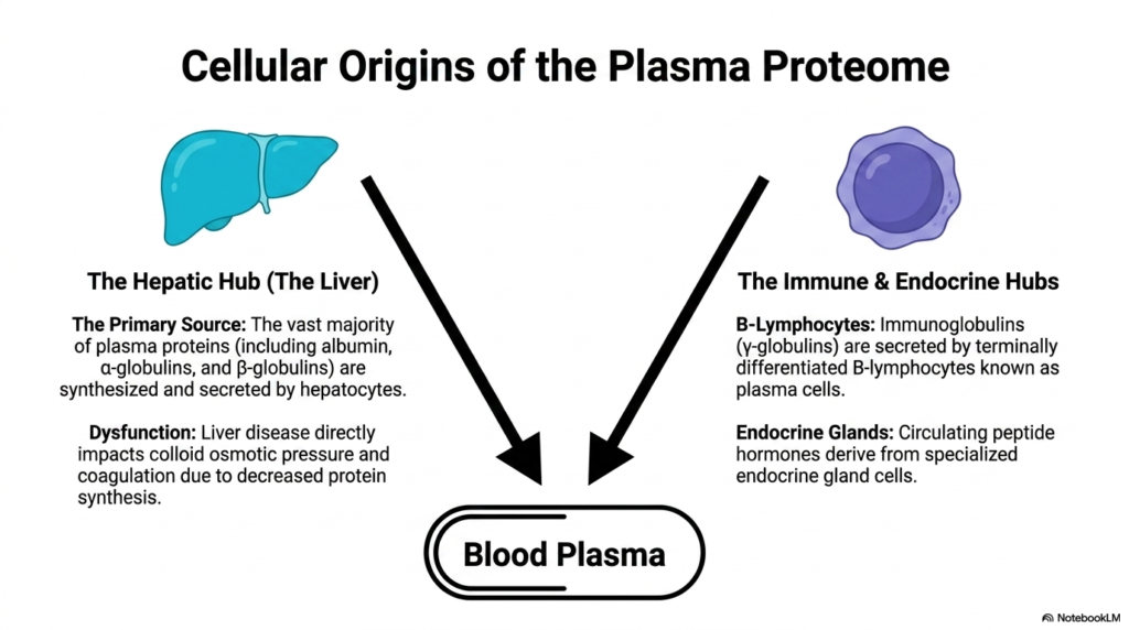
Slide eight visually and biochemically details the distinct cellular origins of the circulating proteome, explaining exactly where the body manufactures these vital plasma proteins. The slide perfectly illustrates a dual-origin system, sharply dividing synthesis between the liver’s hepatic hub and specialized immune and endocrine hubs distributed throughout the body. Understanding this geographical origin is critical for medical students, as organ failure directly correlates with very specific, measurable protein deficiencies in clinical laboratory tests.
The liver is the primary source for synthesizing plasma proteins. Hepatocytes, the specialized functional cells of the liver, relentlessly manufacture the vast majority of the circulating proteome. This immense biological factory is solely responsible for producing all circulating albumin, alongside nearly all alpha and beta globulins. Because of this centralized production, any significant hepatic dysfunction, such as cirrhosis or severe hepatitis, immediately results in a catastrophic drop in total protein synthesis, severely disrupting human fluid dynamics and coagulation pathways.
When the liver fails to produce sufficient plasma proteins, the entire body suffers. The loss of albumin immediately destroys the delicate colloid osmotic pressure, leading to systemic fluid retention and severe abdominal swelling. Simultaneously, the drastic reduction in alpha and beta globulins deprives the blood of vital coagulation factors, leaving the patient extremely vulnerable to uncontrolled bleeding. This direct cause-and-effect relationship perfectly highlights why clinicians use comprehensive protein panels as a primary diagnostic window into overall liver health and functional capacity.
In stark contrast, the gamma globulin fraction of plasma proteins is synthesized completely outside the liver. These specialized soluble antibodies are exclusively secreted by terminally differentiated B-lymphocytes, biologically known as plasma cells, representing the immune hub. Furthermore, circulating peptide hormones are derived from specialized endocrine gland cells. This crucial separation of origin explains why patients with severe liver failure can still maintain a highly robust, fully functional adaptive immune system, as antibody production remains completely independent of hepatic function.
Slide 9: Molecular Aging and the Clearance of Plasma Proteins
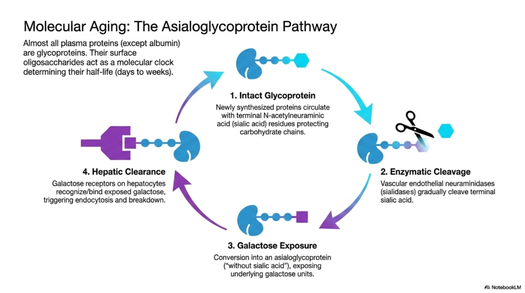
Slide nine delves into the fascinating asialoglycoprotein pathway, revealing the precise molecular clock that governs the lifespan and molecular aging of circulating plasma proteins. With the notable exception of albumin, almost all of these functional macromolecules are heavily structured glycoproteins. Their exterior surfaces are decorated with complex oligosaccharides that completely dictate their physiological half-life, which typically ranges from several days to a few weeks. This slide maps the step-by-step enzymatic degradation process that safely removes old molecules from circulation.
The lifecycle begins with an intact, newly synthesized glycoprotein entering the bloodstream. These fresh plasma proteins circulate safely because their delicate carbohydrate chains are completely capped and protected by terminal N-acetylneuraminic acid (sialic acid) residues. These specific sialic acid shields prevent the liver from prematurely recognizing and destroying the valuable functional molecules. As long as this chemical cap remains completely intact, the protein is free to continually execute its specialized biological duties throughout the vascular system.
However, the vascular environment contains specific endothelial enzymes known as neuraminidases, or sialidases. Over time, these enzymes gradually cleave the terminal sialic acid caps off circulating plasma proteins. This enzymatic cleavage acts as a biological timer. Once the sialic acid is entirely removed, the molecule undergoes a chemical conversion into an asialoglycoprotein, literally translating to a glycoprotein entirely without sialic acid. This crucial conversion suddenly exposes the underlying, previously hidden galactose carbohydrate units to the surrounding bloodstream.
The final stage involves strict hepatic clearance. Specialized galactose receptors located on the surface of liver hepatocytes instantly recognize and bind to the newly exposed galactose residues on these aged plasma proteins. This targeted binding rapidly triggers endocytosis, pulling the expired molecule directly into the liver cell for complete chemical breakdown and recycling. Medical students must thoroughly understand this elegant clearance pathway, as it brilliantly demonstrates how the human body strictly manages protein turnover without wasting valuable amino acid resources.
Slide 10: Dynamic Responses to Pathology Involving Plasma Proteins
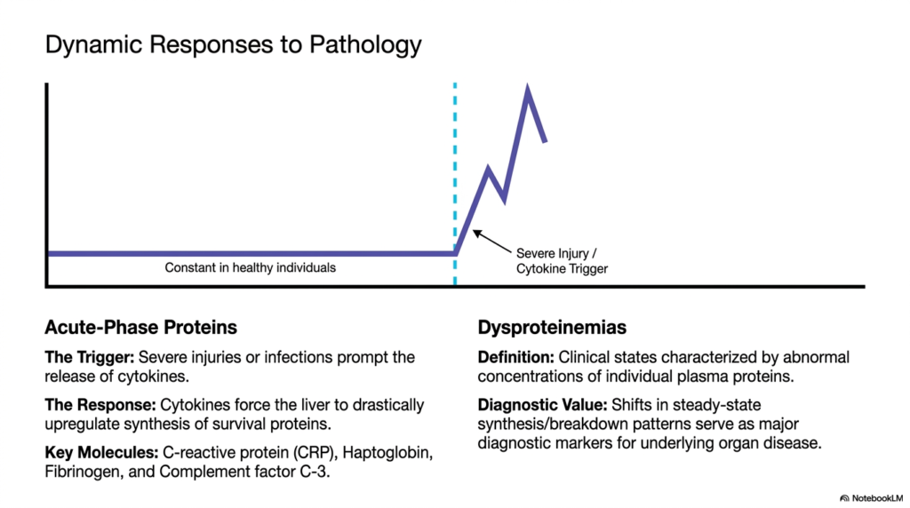
Slide ten illustrates the intense dynamic responses of plasma proteins during states of clinical pathology, moving away from healthy baseline metrics into the realm of diagnostic disease tracking. The prominent line graph on the slide visually depicts a stark, sudden shift in protein synthesis triggered by severe injury or active infection. In perfectly healthy individuals, protein concentrations remain remarkably constant. However, the moment physiological trauma occurs, the biochemical landscape drastically and violently shifts to ensure systemic survival.
The left side of the slide introduces the critical concept of acute-phase proteins. When severe physical injuries or aggressive infections occur, the body instantly releases powerful chemical messengers called cytokines. These specific cytokines travel directly to the liver, forcing the hepatic hub to drastically and immediately upregulate the synthesis of vital survival molecules. The human body strategically prioritizes the rapid mass production of these specific plasma proteins to aggressively combat systemic inflammation and immediately stabilize severely damaged vascular tissues.
Key acute-phase plasma proteins include vital defensive and structural molecules like C-reactive protein, haptoglobin, fibrinogen, and complement factor C-3. The massive spike in C-reactive protein, in particular, serves as one of the most reliable and widely used clinical indicators of severe systemic inflammation in modern medicine. By tracking the rapidly rising and falling blood concentrations of these specific survival molecules, clinicians can accurately measure the severity of an infection and monitor a patient’s response to ongoing medical treatments.
The right side of the slide defines dysproteinemias, which are clinical states explicitly characterized by highly abnormal concentrations of individual plasma proteins. These dangerous diagnostic shifts in steady-state synthesis and breakdown patterns serve as major clinical markers for underlying organ disease or bone marrow cancers. Understanding how these protein levels dynamically react to various pathologies allows medical students to effectively read a patient’s internal biochemical state, transforming abstract numerical laboratory results into clear, actionable, and life-saving clinical diagnoses.
Slide 11: Carrier Electrophoresis Principles for Plasma Proteins
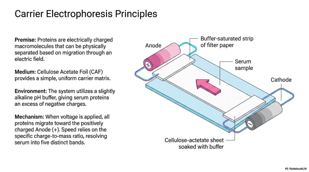
Slide eleven breaks down the mechanical and chemical principles behind carrier electrophoresis, the fundamental laboratory technique used to isolate and analyze plasma proteins. The underlying premise is that proteins are highly complex, electrically charged macromolecules. Because of their distinct surface charges and varied molecular weights, these molecules can be physically and reliably separated from one another simply by forcing them to migrate through a controlled, active electric field within a laboratory setting.
The structural medium illustrated on this slide is cellulose acetate foil. This specific material provides an incredibly simple, highly uniform carrier matrix for the chemical separation process. To manipulate the plasma proteins, the entire system utilizes a strictly regulated, slightly alkaline pH buffer. This specific alkaline environment is absolutely crucial because it forces all the serum proteins to accept an excess of negative electrical charges. This universal negative charge ensures that every molecule will predictably move in the exact same physical direction.
When a high electrical voltage is applied to the buffer-saturated system, the fundamental mechanism of migration begins. All the negatively charged plasma proteins immediately begin actively migrating toward the positively charged Anode. However, they do not travel together. Their individual movement speed completely relies on their specific, inherent charge-to-mass ratio. Small, highly negatively charged molecules sprint rapidly across the cellulose matrix, while massive, weakly charged molecules struggle to move forward, effectively separating the mixture.
This precise mechanism reliably and consistently resolves raw human serum into five highly distinct visual bands. By mastering the fundamental physics and biochemical environments depicted in this slide, medical students easily comprehend exactly how invisible, microscopic molecular structures can be aggressively manipulated for clear diagnostic visualization. Understanding the distinct behavior of plasma proteins within an electrical field is essential for correctly interpreting the final clinical gels used daily in modern diagnostic laboratories worldwide.
Slide 12: From Migration to Measurement of Plasma Proteins
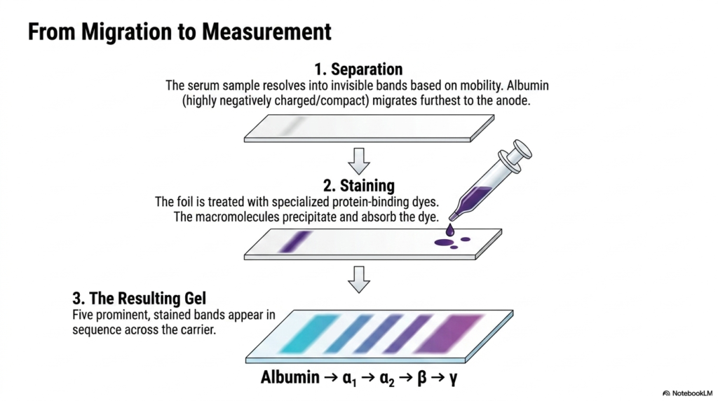
Slide twelve visually walks the student through the exact, step-by-step laboratory procedure that transforms invisible molecular migration into a clearly measurable diagnostic gel for plasma proteins. The graphic simplifies the highly complex electrophoresis process into three easily understandable stages: separation, staining, and the final resulting gel. This practical sequence bridges the gap between the theoretical physics discussed previously and the actual physical artifact that a clinical biochemist or medical physician examines when formally diagnosing a patient’s specific metabolic state.
The first critical step involves the active physical separation of the raw serum sample. As the electrical current runs, the transparent plasma proteins resolve into distinct bands based entirely on their individual electrophoretic mobility. Albumin, being a highly compact and heavily negatively charged macromolecule, migrates the farthest distance toward the positive anode. At this specific stage, the cellulose acetate foil appears totally blank to the naked eye, necessitating a powerful chemical intervention to reveal the hidden molecular data.
The second vital step is the chemical staining process. The delicate foil is carefully removed from the electrical field and aggressively treated with highly specialized protein-binding dyes. When these powerful dyes completely wash over the matrix, the isolated macromolecules instantly precipitate and heavily absorb the dark pigment. This crucial chemical reaction permanently locks the separated plasma proteins directly in place and transforms their invisible molecular presence into a deeply colored, highly visible physical stain across the thin carrier foil.
The final resulting gel clearly reveals five prominent, deeply stained bands spanning in sequence directly across the carrier medium. From the fastest migrating molecule to the absolute slowest, the established diagnostic order is always Albumin, followed by alpha-1, alpha-2, beta, and finally the gamma globulins. Recognizing this exact standard distribution pattern of plasma proteins is an absolutely fundamental skill for any medical student, as deviations from this strict visual sequence immediately indicate severe, potentially fatal underlying clinical diseases.
Slide 13: Quantitative Assessment of Plasma Proteins via Densitometry
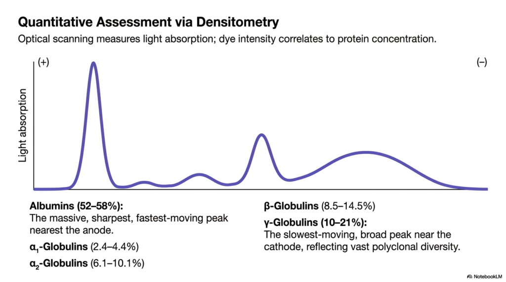
Slide thirteen introduces the critical concept of quantitative densitometry, demonstrating exactly how modern clinicians accurately measure the specific concentrations of separated plasma proteins. While a stained electrophoresis gel provides a great qualitative visual, modern medicine requires precise, exact numerical data. This is achieved through highly advanced optical scanning technology, which systematically measures light absorption directly across the stained foil. The fundamental principle is remarkably simple: the dark intensity of the chemical dye perfectly correlates to the total protein concentration in that exact location.
The massive graph displayed on the slide represents the optical scanner’s final readout, mapping light absorption against the physical migration distance. The most striking feature is the enormous, razor-sharp peak located nearest the positive anode. This massive peak explicitly represents the albumin fraction, normally comprising an astonishing fifty-two to fifty-eight percent of the total mixture. Because albumin consists of purely identical, non-glycosylated molecules, it moves as a unified, massive wave, creating this distinct, rapidly moving, and extremely tall optical peak.
Following the primary peak, the scanner detects the smaller, distinct elevations representing the various globulin fractions. The alpha-1 fraction accounts for roughly two to four percent, followed by alpha-2 at six to ten percent, and the beta globulins reaching up to fourteen percent. These specific plasma proteins create slightly wider, shorter peaks because they contain a wider, more diverse mixture of differently sized, heavily glycosylated transport and regulatory molecules, spreading out slightly more during their slower electrical migration through the cellulose acetate matrix.
Finally, the optical scanner maps the gamma globulin fraction, residing nearest the negative cathode. This specific grouping typically represents ten to twenty-one percent of the total plasma proteins. Crucially, the gamma peak is always exceptionally broad and visually flattened compared to albumin. This distinct shape directly reflects the vast polyclonal diversity of human antibodies, which possess wildly different masses and slight electrical charge variations. Mastering this densitometry curve allows students to instantly identify dangerous monoclonal spikes or severe molecular deficiencies in patients.
Slide 14: The Gamma-Globulin Fraction of Plasma Proteins
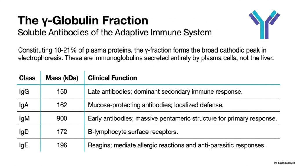
Slide fourteen focuses entirely on the gamma-globulin fraction, the incredibly diverse subset of plasma proteins completely dedicated to the human body’s advanced adaptive immune system. Constituting ten to twenty-one percent of the total circulating proteome, this specific fraction forms the uniquely broad, slowly moving cathodic peak prominently seen during clinical electrophoresis. Unlike all other major fractions discussed previously, these powerful immunoglobulins are completely independent of the liver, being synthesized and secreted exclusively by highly specialized immune cells called plasma cells.
The table provided on the slide breaks down the five major classes of these defensive plasma proteins, commonly known as antibodies. The most abundant class is Immunoglobulin G, massive 150-kilodalton molecules that act as late-stage antibodies. They absolutely dominate the secondary immune response, providing powerful, long-lasting systemic immunity against previously encountered biological pathogens. Following this, Immunoglobulin A, weighing one hundred and sixty-two kilodaltons, serves a highly specialized localized defense role, specifically protecting the body’s vulnerable mucosal membranes from aggressive external infections.
Immunoglobulin M is a massive, incredibly powerful pentameric structure weighing an astonishing nine hundred kilodaltons. Due to its massive size and multiple binding sites, it serves as the absolute first line of adaptive defense, providing an immense early primary response before the body formally manufactures targeted IgG. Immunoglobulin D, weighing 172 kilodaltons, functions primarily as a crucial surface receptor directly on B-lymphocytes, helping these vital immune cells correctly identify and lock onto dangerous foreign antigens circulating within the blood.
Finally, Immunoglobulin E, weighing 196 kilodaltons, acts as the primary molecular mediator for powerful allergic reactions and aggressive anti-parasitic biological responses, commonly known as reagins. By understanding the distinct mass and specific clinical function of each antibody class, medical students can fully comprehend the immense complexity of the gamma fraction. These specialized plasma proteins represent the pinnacle of evolutionary defense, showcasing how highly targeted, soluble molecular structures actively keep the human body completely safe from microscopic annihilation.
Slide 15: The Plasma Protein Matrix: High-Yield Summary
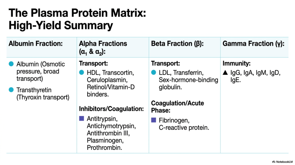
The final slide, slide fifteen, serves as the ultimate high-yield summary, meticulously organizing the vast complexities of plasma proteins into a single, easily digestible matrix. This comprehensive table serves as a vital study tool for medical students, perfectly aligning the four major electrophoretic fractions with their overarching clinical and physiological functions. By consolidating this immense amount of biochemical data, the slide solidifies the core concepts of molecular transport, complex coagulation pathways, strict enzymatic inhibition, and advanced adaptive immunity discussed throughout the presentation.
The matrix elegantly reiterates the absolute dominance of the albumin fraction. As explicitly stated, these specific plasma proteins are entirely responsible for maintaining broad vascular transport and generating life-sustaining osmotic pressure. It correctly lists transthyretin alongside albumin, ensuring students remember its specific role in thyroxin transport. Moving laterally, the alpha fractions are perfectly summarized by their dual roles. They handle the specialized transport of critical lipids and vitamins via HDL and ceruloplasmin, while simultaneously providing vital enzymatic inhibitors and coagulation precursors like antitrypsin and prothrombin.
The beta fraction summary reinforces its massive importance in both heavy transport and the acute-phase inflammatory response. It highlights vital plasma proteins such as low-density lipoproteins for fat transport and transferrin for iron distribution, alongside crucial reactive molecules like fibrinogen and C-reactive protein. This perfectly encapsulates how the beta globulins bridge the gap between daily metabolic maintenance and violent, immediate reactions to severe physical trauma. The clear categorization helps students rapidly connect specific molecular structures to their corresponding, highly critical physiological tasks.
Finally, the matrix isolates the gamma fraction, reaffirming its exclusive dedication to human immunity. By explicitly listing the specific immunoglobulins, the table ensures future clinicians perfectly separate these defensive molecules from the liver-synthesized components. Ultimately, this final high-yield summary perfectly encapsulates the incredible, life-sustaining nature of plasma proteins. It successfully transforms hours of complex, dense biochemical study into a highly structured, logical, and deeply interconnected biological system that students can confidently apply directly to future clinical diagnostic practices.
Please read our Content Disclaimer Statement.
Check out our social media channels: