138. Neurotransmitter Receptors: How Synapses Receive and Regulate Chemical Signals
Have you ever wondered how your brain converts electrical impulses into thoughts, movements, and emotions in a fraction of a second? The secret lies at the synapse, where chemical messengers bridge the cellular gap to deliver crucial instructions. To receive these chemical signals, cells rely on specialized membrane proteins that translate chemical binding into cellular action. This slide deck provides a comprehensive breakdown of these essential cellular gateways and their biochemical pathways. Designed for college and medical students, this visual guide explores how these molecular structures operate, classify, synthesize, and terminate signals across human physiological systems.
Slide 1: Introduction to Neurotransmitter Receptors and Metabolism
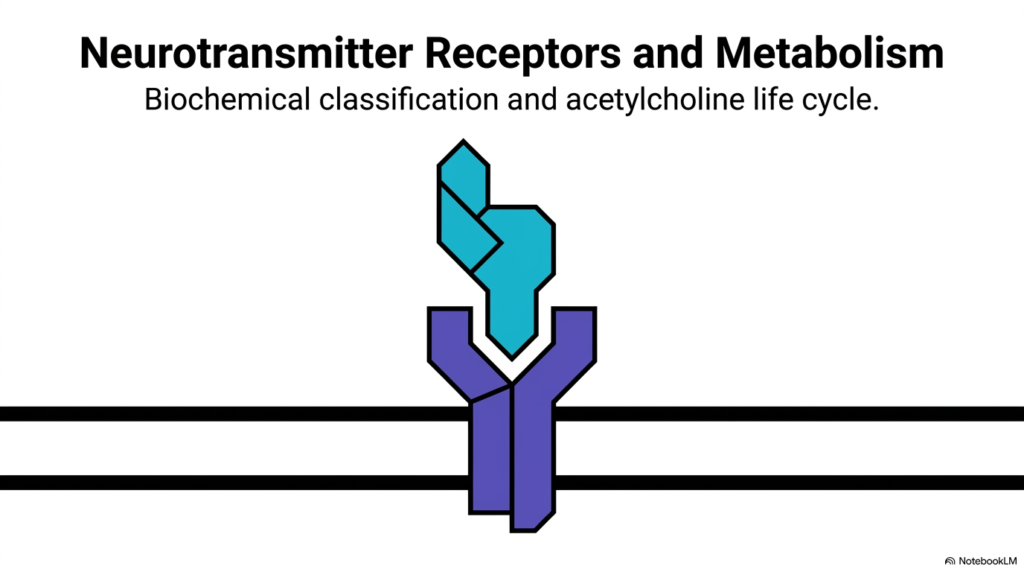
The human nervous system depends entirely on chemical communication to control every physiological function across the body. At the center of this communication network are Neurotransmitter Receptors, specialized protein gateways embedded in target cell membranes. When a presynaptic neuron releases chemical messengers into the synaptic gap, these Neurotransmitter Receptors selectively bind the signaling molecules to initiate an immediate downstream response. Without these dedicated membrane proteins, chemical signals would drift aimlessly through the extracellular space without triggering cellular activity.
Beyond basic signal recognition, studying Neurotransmitter Receptors requires analyzing the complete metabolic life cycle of the chemical ligands that activate them. Acetylcholine serves as the classic prototype for this fundamental biological process. A complete understanding demands examining how the signaling ligand is synthesized inside the presynaptic terminal, packaged into storage vesicles, released across the synaptic cleft, and rapidly cleared after binding.
This opening slide introduces both structural classification and metabolic life cycles for students. College and medical students will learn how rapid channel opening and complex biochemical cascades work together to maintain total physiological harmony across neural circuits. As we advance through this deck, keep in mind that Neurotransmitter Receptors are the ultimate decision-makers at the synapse, dictating whether a signal continues or stops.
Slide 2: The Postsynaptic Gateway of Neurotransmitter Receptors
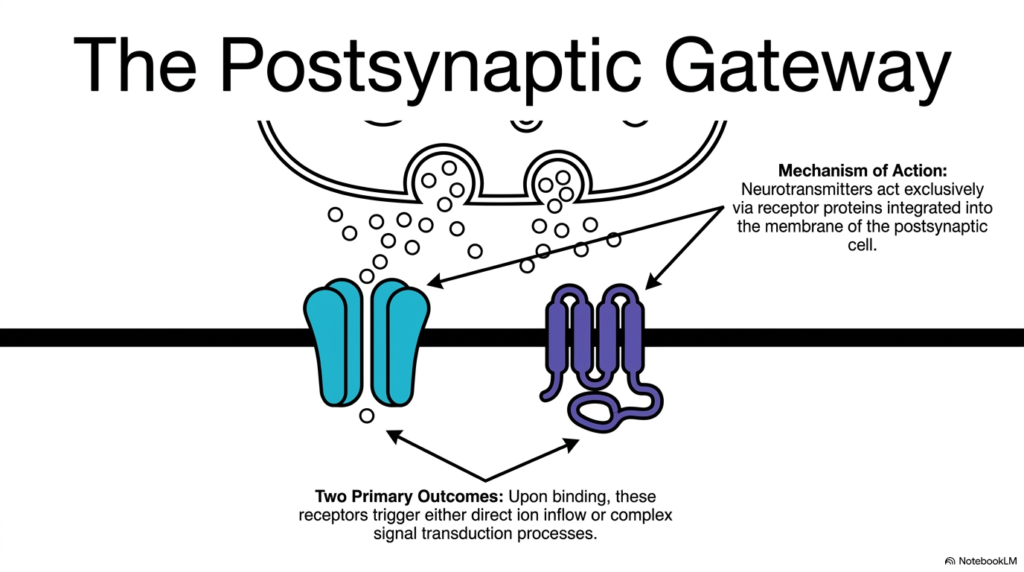
Synaptic transmission relies on a highly organized structural architecture to convert extracellular chemical signals into precise intracellular responses. Integrated directly into the postsynaptic lipid bilayer, neurotransmitter receptors serve as the primary molecular targets for released chemical messengers. When neurotransmitters diffuse across the narrow synaptic cleft, they interact exclusively with these membrane-bound proteins. This exclusive interaction guarantees that circulating signaling molecules produce specific, localized effects on target cells rather than causing non-specific activation across neighboring tissues.
Upon ligand binding, Neurotransmitter Receptors drive cellular communication through two distinct physiological outcomes. The first outcome involves direct ion inflow, where channel activation alters membrane potential in milliseconds to excite or inhibit the postsynaptic cell. The second outcome triggers complex signal transduction processes, initiating enzyme cascades that regulate intracellular second messengers over longer timeframes.
Understanding these dual mechanisms reveals how postsynaptic membranes process incoming chemical messages. By utilizing different classes of Neurotransmitter Receptors, a single chemical signal can produce either a rapid electrical spike or a prolonged metabolic change, giving the central nervous system remarkable computational flexibility. As students analyze these cellular gateways, appreciating how localized membrane integration prevents generalized signal decay across neural circuits remains essential for mastering fundamental neurophysiology in medical education.
Slide 3: Structural Classification of Neurotransmitter Receptors
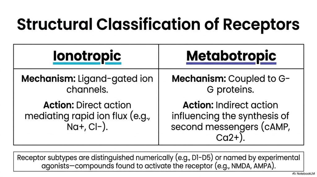
To understand how chemical messages produce diverse biological actions, biochemists categorize Neurotransmitter Receptors into two main structural classes: ionotropic and metabotropic. Ionotropic receptors are ligand-gated ion channels that combine the transmitter binding site and the channel pore into a single transmembrane protein complex. When a ligand attaches to the extracellular domain, the channel undergoes a rapid conformational change that allows ion flux across the postsynaptic membrane. This direct action mediates fast synaptic transmission within milliseconds, driving rapid processes like skeletal muscle contraction and immediate reflex pathways.
In contrast, metabotropic Neurotransmitter Receptors mediate indirect, slower responses by coupling to intracellular G-proteins. Instead of containing an integral ion channel, these receptor proteins activate enzymatic cascades that modulate second messengers such as cyclic AMP or calcium ions. This indirect mechanism lets a single ligand-binding event amplify signals throughout the cell interior over seconds or minutes.
Within these broad structural categories, specific Neurotransmitter Receptors are further distinguished numerically, such as dopamine D1 through D5, or named after experimental agonists like NMDA and AMPA that selectively activate them in laboratory research. Recognizing these structural distinctions allows college students to predict whether a receptor will trigger fast electrical spikes or slow metabolic modulation during neuronal activity.
Slide 4: Ionotropic Dynamics of Neurotransmitter Receptors
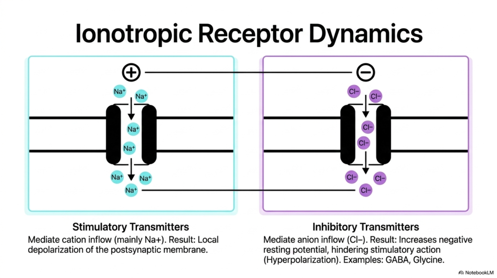
Ionotropic Neurotransmitter Receptors operate as specialized transmembrane channels that open directly when bound by specific signaling molecules. The functional outcome of ionotropic activation depends on whether the receptor channel selectively conducts positively charged cations or negatively charged anions. Stimulatory Neurotransmitter Receptors mediate the inward flux of cations, primarily sodium ions. This influx of positive charge reduces the negative electrical potential across the postsynaptic membrane, causing local depolarization. When this local depolarization reaches a critical threshold, it triggers an action potential that propagates along the postsynaptic neuron.
Conversely, inhibitory Neurotransmitter Receptors mediate the inward flux of anions, specifically chloride ions. The entering chloride ions make the resting potential inside the cell more negative, causing hyperpolarization that hinders excitatory electrical activity. Major inhibitory transmitters like gamma-aminobutyric acid and glycine act through these anion-selective channels to prevent over-excitation in neural networks.
By balancing cation-mediated excitation with anion-mediated inhibition, ionotropic Neurotransmitter Receptors maintain precise control over cellular excitability throughout the central nervous system. This balance keeps electrical signals within safe functional boundaries during rapid neural processing, protecting delicate brain circuits from dangerous hyper-excitability while enabling precise millisecond communication across complex cortical pathways and peripheral nerve circuits in human physiology and neurobiology coursework for medical education.
Slide 5: Signal Transduction by Metabotropic Neurotransmitter Receptors
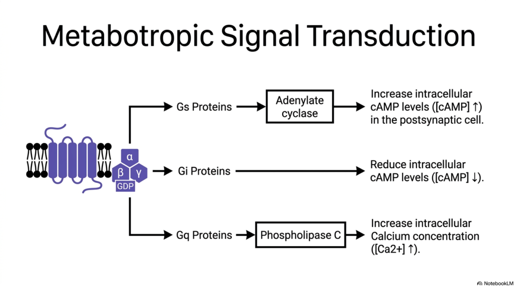
Metabotropic Neurotransmitter Receptors alter cellular physiology through multi-step signal transduction cascades mediated by heterotrimeric G-proteins. When a ligand binds to the extracellular domain of these seven-transmembrane receptors, the associated G-protein complex exchanges bound GDP for GTP, causing the G-protein subunit to dissociate. The downstream functional outcome depends on the specific G-protein alpha subunit coupled to the receptor. For instance, Neurotransmitter Receptors linked to Gs proteins activate adenylate cyclase, which converts ATP into cyclic AMP to increase intracellular second messenger levels and stimulate cellular protein kinases.
Conversely, metabotropic Neurotransmitter Receptors coupled to Gi proteins inhibit adenylate cyclase, reducing intracellular cyclic AMP concentration to dampen cellular activity. A third major pathway involves receptors linked to Gq proteins, which activate phospholipase C to cleave membrane phospholipids into second messengers that elevate intracellular calcium concentrations.
These intracellular signaling pathways allow metabotropic Neurotransmitter Receptors to regulate gene expression, enzyme activity, and membrane channel sensitivity over prolonged timeframes. This G-protein versatility enables long-term cellular adaptation, synaptic plasticity, and complex behavioral modulation throughout human physiology. Medical students must recognize that these enzymatic cascades amplify small extracellular signals into major intracellular changes across diverse organ systems, cellular targets, and clinical medicine contexts across modern healthcare practice and research.
Slide 6: Principal Transmitters and Their Neurotransmitter Receptors
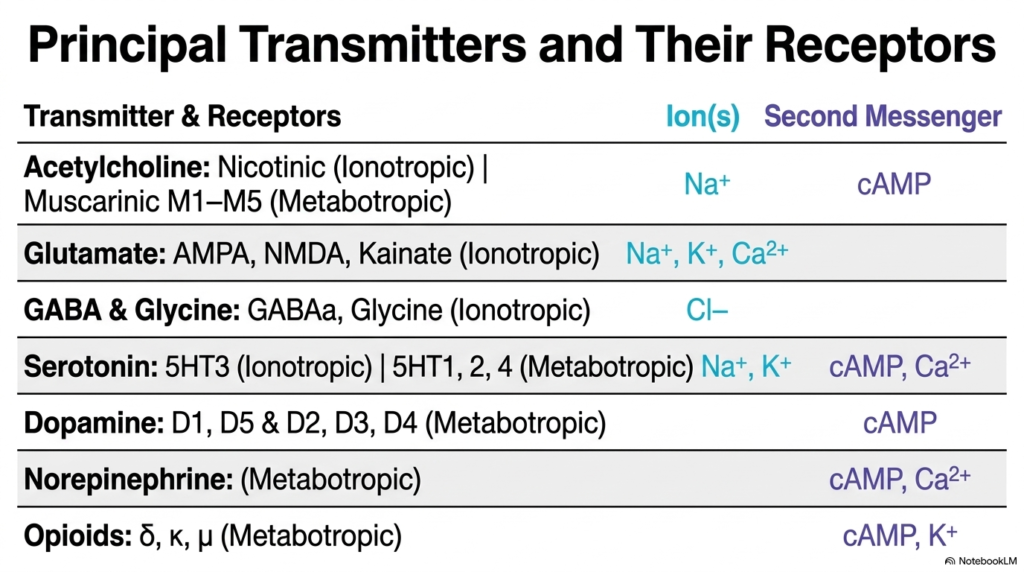
A wide variety of signaling molecules use specialized Neurotransmitter Receptors to coordinate physiological functions across the body. Acetylcholine activates both ionotropic nicotinic channels, which drive sodium influx, and metabotropic muscarinic M1-M5 subtypes, which alter cyclic AMP levels. Glutamate is the primary excitatory neurotransmitter in the central nervous system, acting through ionotropic AMPA, NMDA, and kainate Receptors to allow the flow of sodium, potassium, and calcium ions required for synaptic plasticity and memory formation.
In contrast, major inhibitory transmission relies on gamma-aminobutyric acid and glycine, which activate ionotropic Neurotransmitter Receptors that conduct chloride ions to hyperpolarize postsynaptic cells. Biogenic amines exhibit diverse functional profiles; serotonin acts on ionotropic 5-HT3 channels as well as metabotropic 5-HT1, 2, and 4 subtypes. Dopamine utilizes metabotropic D1 through D5 receptors to modulate cyclic AMP, while norepinephrine acts exclusively through metabotropic pathways to regulate cyclic AMP and intracellular calcium.
Finally, opioid transmitters bind delta, kappa, and mu metabotropic receptors to modulate cyclic AMP and potassium conductance to regulate pain. Mapping these major signaling chemicals to their corresponding Neurotransmitter Receptors illustrates how diverse biological functions are controlled across human tissues, providing an essential biochemical reference for college and medical student examination review and coursework.
Slide 7: Discovery and Subtypes of Acetylcholine Neurotransmitter Receptors
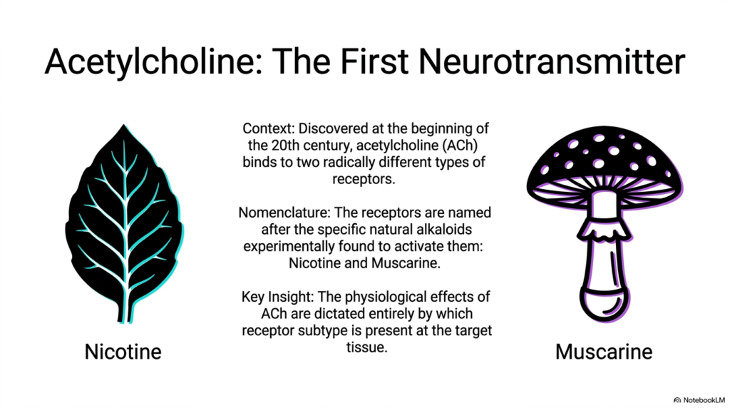
Discovered in the early twentieth century, acetylcholine was the first chemical messenger identified in human physiology. Early pharmacological research revealed that acetylcholine does not produce uniform responses in all tissues; instead, it binds to two distinct classes of Neurotransmitter Receptors. To distinguish these receptor populations, researchers used natural plant alkaloids that selectively mimicked acetylcholine’s action at specific tissue sites. This experimental strategy led to the modern classification of cholinergic receptors based on their sensitivity to plant toxins.
The two primary classes of acetylcholine Neurotransmitter Receptors were named after nicotine, derived from tobacco leaves, and muscarine, isolated from the poisonous mushroom Amanita muscaria. Pharmacologists discovered that nicotine selectively stimulates fast electrical responses in skeletal muscle and autonomic ganglia, whereas muscarine mimics acetylcholine actions on visceral smooth muscle and cardiac tissue.
The key physiological insight from this discovery is that acetylcholine’s biological effect depends entirely on which receptor subtype is present on the target membrane. These acetylcholine neurotransmitter receptors show how target tissues customize chemical responses using distinct receptor proteins, establishing a foundational principle in pharmacology for developing selective drugs that target specific tissue responses without affecting non-target organ systems across human anatomy and clinical practice.
Slide 8: Comparing Nicotinic and Muscarinic Neurotransmitter Receptors
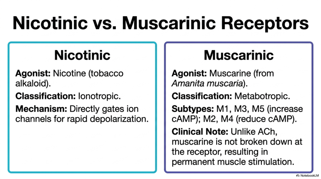
Comparing nicotinic and muscarinic subtypes highlights how structural differences determine functional roles among Neurotransmitter Receptors. Nicotinic receptors are ionotropic ligand-gated channels activated by the tobacco alkaloid nicotine. Upon acetylcholine binding, nicotinic channels open directly to allow rapid sodium influx, producing immediate membrane depolarization at neuromuscular junctions and autonomic ganglia. This rapid gating makes nicotinic Neurotransmitter Receptors ideal for physiological processes requiring instantaneous communication, such as voluntary muscle contraction and rapid sensory reflex arcs across peripheral nerve pathways.
In contrast, muscarinic receptors are metabotropic G-protein-coupled structures activated by the fungal toxin muscarine. Muscarinic Neurotransmitter Receptors are divided into distinct functional subtypes: M1, M3, and M5 increase intracellular cyclic AMP or calcium, while M2 and M4 reduce cyclic AMP levels.
A critical clinical insight involves agonist clearance; unlike acetylcholine, muscarine is not degraded by acetylcholinesterase at the receptor site. When muscarine binds to muscarinic Neurotransmitter Receptors, it causes permanent, uncontrolled receptor stimulation, leading to severe autonomic distortion, continuous smooth muscle spasm, and profuse secretory output during mushroom poisoning. This striking contrast reinforces how receptor structure governs both normal human physiology and toxicological pathology in clinical medicine, emergency healthcare, and modern pharmacology curricula across medical schools, pharmacology departments, and clinical training programs.
Slide 9: Synthesis and Storage of Ligands for Neurotransmitter Receptors
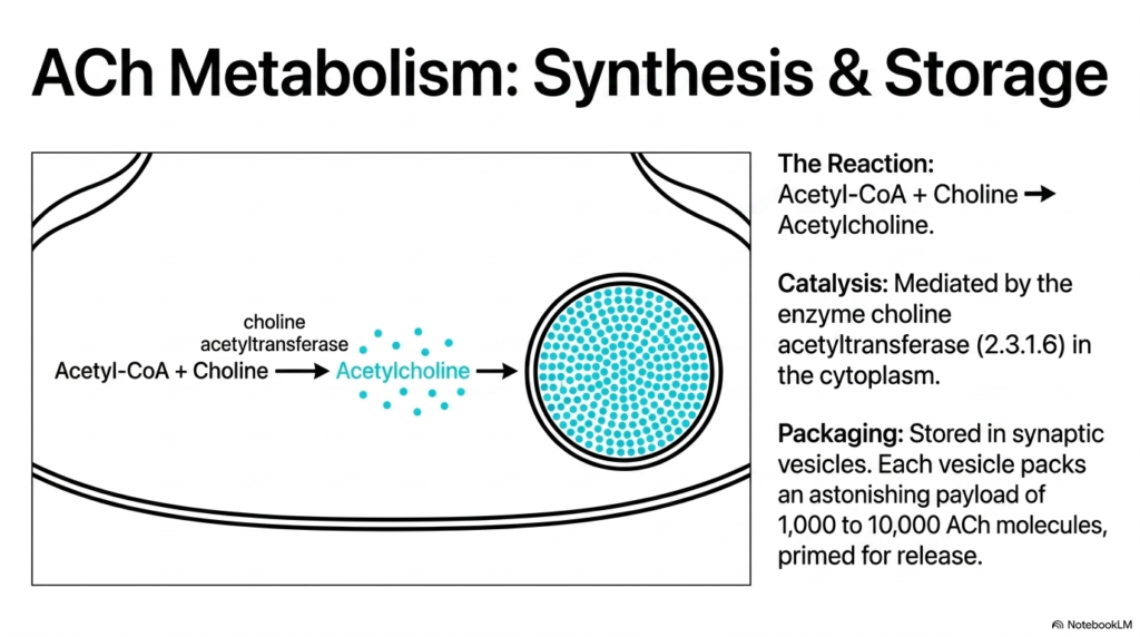
For postsynaptic Neurotransmitter Receptors to function reliably, the presynaptic neuron must maintain a continuous supply of chemical signaling molecules. Acetylcholine is synthesized in the neuronal cytoplasm through a single enzymatic reaction that combines acetyl-CoA and choline. The cytoplasmic enzyme choline acetyltransferase catalyzes this condensation reaction, classified under enzyme commission number 2.3.1.6. Mitochondrial respiration supplies acetyl-CoA, while specialized membrane transport proteins import choline into the nerve terminal from the extracellular fluid.
Once synthesized in the cytoplasm, acetylcholine molecules must be concentrated into storage organelles to prevent premature breakdown and ensure organized release toward postsynaptic Neurotransmitter Receptors. Active vesicular transporters pump acetylcholine into specialized synaptic vesicles anchored near the active zone of the presynaptic membrane. Each individual vesicle packs an astonishing payload containing between one thousand and ten thousand acetylcholine molecules.
This dense vesicular storage ensures that incoming action potentials release concentrated packets of chemical messenger, providing the threshold ligand concentration required to rapidly open postsynaptic Neurotransmitter Receptors during synaptic transmission. Without this efficient metabolic packaging, chemical communication across the synaptic cleft would fail to activate target-cell pathways, disrupting physiological signaling throughout neural networks, brain regions, and peripheral effector organs.
Slide 10: Release and Action of Ligands on Neurotransmitter Receptors
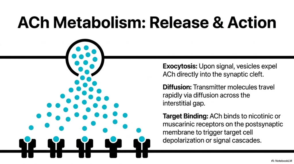
When an electrical action potential invades the presynaptic terminal, voltage-gated calcium channels open, triggering regulated exocytosis of synaptic vesicles. The storage vesicle membrane fuses with the presynaptic plasma membrane, releasing thousands of acetylcholine molecules directly into the synaptic cleft. Once inside the interstitial gap, the released transmitter molecules travel rapidly via simple diffusion toward the postsynaptic cell. The narrow width of the synaptic cleft ensures that chemical diffusion occurs in microseconds, minimizing signal delay between neurons.
Upon reaching the postsynaptic membrane, acetylcholine molecules bind selectively to localized Neurotransmitter Receptors. Depending on the target cell type, the ligand binds to either nicotinic ion channels to trigger immediate sodium influx and cell depolarization, or muscarinic G-protein structures to initiate intracellular signal cascades.
This selective binding step is the central event of chemical transmission, transforming fluid chemical diffusion into a specific electrical or metabolic signal. By interacting with these postsynaptic Neurotransmitter Receptors, acetylcholine successfully delivers the neural command across the synaptic barrier to drive targeted cellular responses. Understanding this dynamic interaction helps medical students grasp how presynaptic exocytosis directly dictates postsynaptic electrical and metabolic outcomes, highlighting how Neurotransmitter Receptors govern physiological communication across chemical synapses throughout human physiology.
Slide 11: Enzymatic Hydrolysis to Reset Neurotransmitter Receptors
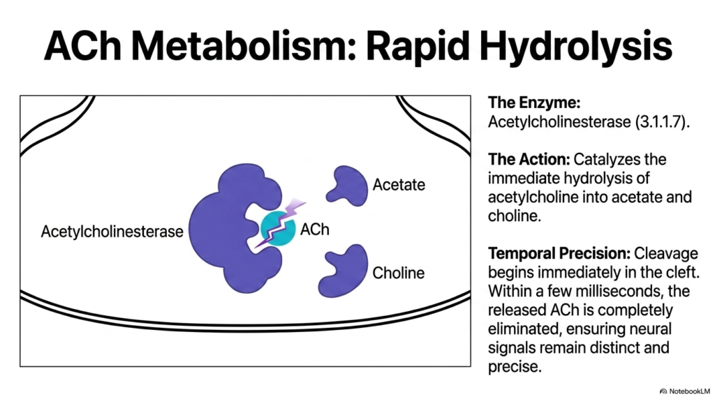
After acetylcholine binds to postsynaptic Neurotransmitter Receptors, the chemical signal must be terminated rapidly to prevent continuous receptor stimulation and allow subsequent neural messages to be received. Signal termination at cholinergic synapses relies on the enzyme acetylcholinesterase, systematically designated as enzyme commission number 3.1.1.7. Positioned directly within the synaptic cleft and anchored to the postsynaptic basement membrane, acetylcholinesterase catalyzes the immediate hydrolysis of acetylcholine into two inactive metabolites: acetate and choline.
This enzymatic cleavage is temporally precise, beginning the instant acetylcholine enters the cleft. Within a few milliseconds, acetylcholinesterase eliminates released acetylcholine molecules, freeing the binding sites on postsynaptic Neurotransmitter Receptors. This rapid clearance resets the postsynaptic membrane, preventing receptor desensitization and continuous muscle firing.
By clearing free transmitters in milliseconds, acetylcholinesterase ensures that postsynaptic Neurotransmitter Receptors remain responsive to discrete action potentials. This millisecond timing maintains the high fidelity and temporal accuracy required for precise muscle coordination and rapid neural processing across the human nervous system. Without this rapid enzymatic cleanup, synapses would quickly lock up from persistent, nonstop stimulation of postsynaptic Neurotransmitter Receptors, severely disrupting normal neuromuscular transmission, voluntary muscle tone, and physiological reflex control across central and peripheral neural circuits in human subjects.
Slide 12: Reuptake and Recycling to Sustain Neurotransmitter Receptors
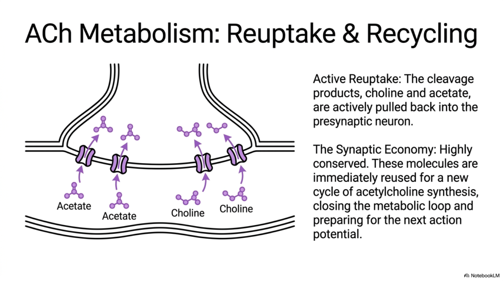
To maintain continuous signal delivery to postsynaptic Neurotransmitter Receptors, neurons rely on efficient reuptake pathways that recycle essential molecular building blocks. Following the rapid hydrolysis of acetylcholine in the synaptic cleft, the resulting cleavage products, choline and acetate, are actively pulled back into the presynaptic terminal. Sodium-dependent high-affinity choline transporters embedded in the presynaptic plasma membrane pump choline from the extracellular space back into the neuronal cytoplasm against its concentration gradient.
This active reuptake mechanism represents a highly conserved synaptic economy within the nervous system. Choline acetyltransferase immediately combines the recovered choline with fresh mitochondrial acetyl-CoA to synthesize new acetylcholine molecules, closing the metabolic loop and preparing the neuron for subsequent action potentials.
Without this active transport recycling system, presynaptic terminals would rapidly deplete their choline reserves during high-frequency firing, leading to failure of chemical transmission at postsynaptic Neurotransmitter Receptors. Recycling cleavage products ensures that target-cell neurotransmitter receptors receive a dependable supply of chemical transmitters during prolonged neural activity, sustaining long-term physiological signaling without cellular fatigue. Thus, metabolic recycling directly supports continuous Neurotransmitter Receptors functionality across active neuronal circuits, autonomic pathways, and somatic motor units throughout human health and physiological performance standards in sports and daily life.
Slide 13: Pharmacological Disruption of Neurotransmitter Receptors
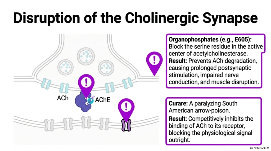
The essential role of cholinergic transmission makes the synapse a prime target for chemical toxins and therapeutic drugs that disrupt Neurotransmitter Receptors or their regulatory enzymes. Organophosphate compounds, such as the pesticide E605, act as irreversible inhibitors by binding to the critical serine residue in the active site of acetylcholinesterase. By blocking enzymatic hydrolysis, organophosphates prevent acetylcholine degradation in the synaptic cleft. This accumulation causes continuous, uncontrolled stimulation of postsynaptic Neurotransmitter Receptors, leading to impaired nerve conduction, violent muscle fasciculations, and fatal respiratory failure.
Conversely, plant toxins can directly target postsynaptic protein sites to block synaptic signaling outright. Curare, a traditional South American arrow poison, acts as a competitive antagonist that selectively binds to nicotinic Neurotransmitter Receptors at the neuromuscular junction. By occupying the agonist binding site without opening the ion channel, curare prevents acetylcholine from binding to its target protein.
This competitive inhibition blocks physiological signal transmission, resulting in flaccid paralysis of skeletal muscles. Studying these pharmacological disruptors shows how interfering with Neurotransmitter Receptors or metabolic enzymes can completely arrest neuromuscular function, providing vital insights for toxicology, emergency medicine, anesthesiology, clinical pharmacology, and comprehensive patient care across healthcare specialties and clinical settings, including hospitals and toxicological units.
Slide 14: The Homeostatic Balance of Neurotransmitter Receptors
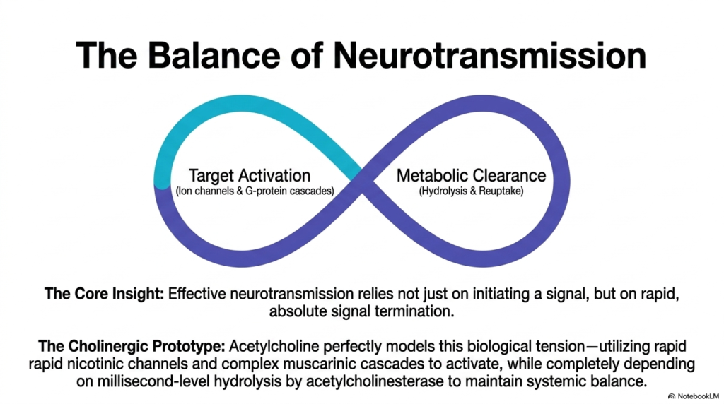
Effective chemical signaling in human physiology requires a perfect homeostatic balance between signal initiation and signal termination. As demonstrated across this slide deck, Neurotransmitter Receptors provide the molecular machinery for target activation through direct ion channels or G-protein cascades. However, initiating a cellular response represents only half of the synaptic equation; absolute signal termination through enzymatic hydrolysis and active reuptake is equally vital for maintaining functional neural circuits.
Cholinergic neurotransmission serves as the prototype for this physiological balance. Acetylcholine utilizes rapid nicotinic channels for immediate depolarization and complex muscarinic cascades for metabolic modulation, while relying entirely on millisecond-level hydrolysis by acetylcholinesterase to reset the synapse. If target activation occurs without rapid clearance, postsynaptic Neurotransmitter Receptors become desensitized, causing signal block and cellular exhaustion.
Conversely, if clearance occurs too rapidly, target proteins fail to reach activation thresholds. Understanding how Neurotransmitter Receptors balance rapid activation with millisecond termination provides college and medical students with a foundational framework for mastering neurobiology and pharmacology across health and disease. In summary, healthy nervous system function depends on balancing activation and clearance at postsynaptic Neurotransmitter Receptors across neural pathways in the human body and peripheral nervous system throughout life and clinical disease states.
Please read our Content Disclaimer Statement.
Check out our social media channels: