80. Lysosomes: Biochemical Structure and Cellular Function
Imagine a bustling city that never sleeps. Without an efficient waste management and recycling plant, trash would accumulate, and the city would collapse. In biology, the cell faces the exact same challenge. The core purpose of this slide deck is to explore the biochemical framework of cellular digestion. Readers will discover the intricate lifecycle, structural anatomy, and crucial pathways that govern these cellular recycling centers, understanding how minor genetic errors can lead to devastating metabolic diseases.
Slide 1: Introduction to Lysosomes and Biochemical Sorting
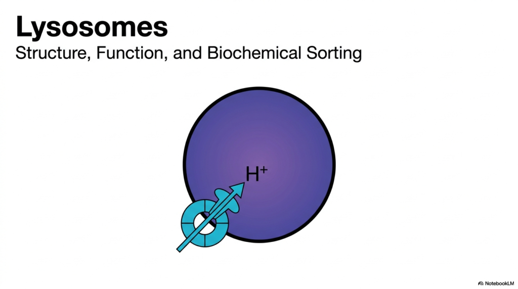
The opening slide introduces the fundamental topic of cellular waste management, focusing squarely on Lysosomes. The visual presents a simplified model of this organelle, highlighting a single, central mechanism: the active pumping of hydrogen ions across a defining membrane. This introductory schematic sets the foundation for understanding how specialized compartments maintain distinct internal environments. Students must recognize that Lysosomes do not merely float passively in the cytoplasm; they are highly dynamic structures that require constant energy expenditure to maintain their unique biochemical identity and fulfill their digestive duties.
Understanding the architecture of Lysosomes is crucial for any medical or biochemistry student. The simple purple sphere depicted in the diagram represents a robust lipid bilayer that serves as a formidable barrier between the destructive payload inside and the delicate cellular machinery outside. The illustration of the proton pump specifically highlights the biochemical sorting and functional readiness that define Lysosomes. Without this physical boundary and active chemical gradient, the very components meant to sustain the cell would indiscriminately dismantle it.
As the slide indicates, the study of Lysosomes extends beyond mere structure to encompass complex biochemical sorting. The cell must meticulously identify, tag, and transport newly synthesized digestive enzymes directly to these organelles. This introductory overview prepares the reader to explore the precise molecular tagging and transport networks required to build and maintain these cellular compartments. Mastery of these sorting pathways reveals how cells achieve remarkable spatial organization, ensuring that the biochemical tools for breakdown reach their correct destination safely and efficiently.
To truly grasp cellular metabolism, one must appreciate the elegant simplicity shown here. The active transport of protons is the single most defining physiological feature that activates the enzymatic core of Lysosomes. This concept will serve as the guiding thread throughout the remaining slides, linking structural biology with energetic cellular demands. The upcoming sections will dissect the exact nature of these enzymes and the intricate journey they take from initial synthesis to final functional maturation within the cell.
Slide 2: The Anatomy of Lysosomes as the Cellular Stomach
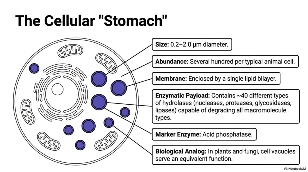
This slide conceptualizes Lysosomes as the cell’s ultimate digestive organ, often referred to as the cellular stomach. Ranging from 0.2 to 2.0 micrometers in diameter, these organelles are enclosed by a single lipid bilayer. A typical animal cell contains several hundred of these vital structures, scattered throughout the cytoplasm. Their sheer abundance underscores the immense, ongoing demand for macromolecule turnover and metabolic recycling. This structural simplicity hides a profound biochemical complexity, as these organelles must safely contain highly destructive chemical reactions.
The defining characteristic of Lysosomes is their potent enzymatic payload. Each vesicle contains roughly 40 distinct acid hydrolases, including nucleases, proteases, glycosidases, and lipases. This versatile chemical arsenal ensures that the cell can efficiently degrade virtually any biological macromolecule, from complex proteins to entire lipid membranes. Acid phosphatase serves as the classic marker enzyme for identifying these compartments in laboratory settings. The presence of such diverse degradative tools highlights the central metabolic role these organelles play in continuous cellular maintenance and adaptation.
It is essential to recognize the evolutionary analogs of Lysosomes across different domains of life. In plant and fungal cells, large structures known as cell vacuoles serve an entirely equivalent function. Whether examining animal cells or plant biology, the core requirement for a specialized, enclosed degradative space remains a universal biological imperative. Students must understand that the compartmentalization of these powerful hydrolases prevents indiscriminate cellular damage, illustrating a perfect balance between metabolic necessity and structural protection.
The concept of the cellular stomach provides an excellent framework for understanding the daily life of a cell. Just as an organism digests food to harvest basic nutrients, the cell relies on Lysosomes to break down larger molecules into their constituent amino acids, sugars, and lipids. These basic building blocks are then exported back into the cytoplasm for reuse. This endless cycle of breakdown and regeneration is the very essence of cellular survival, making the study of these unique organelles absolutely foundational for biochemistry.
Slide 3: Membrane Transport and pH Regulation in Lysosomes
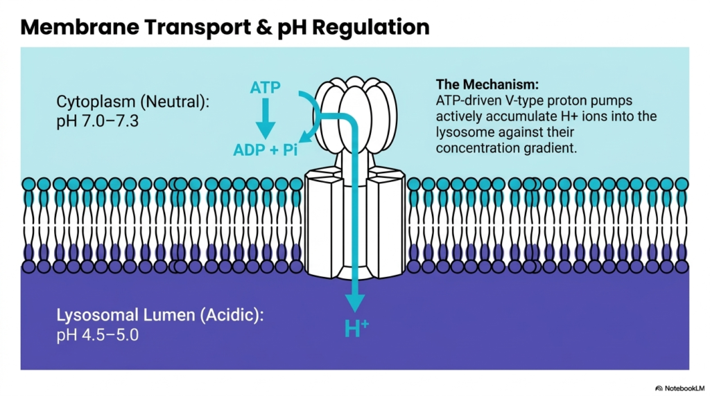
The biochemical magic of Lysosomes relies entirely on strict pH regulation, as detailed in this critical slide. The cytoplasm of a healthy cell maintains a neutral pH ranging from 7.0 to 7.3. In stark contrast, the lumen inside Lysosomes must remain highly acidic, typically at pH 4.5-5.0. This massive concentration gradient does not occur naturally; it requires constant, active intervention by the cell to sustain the harsh environment needed for digestive functions.
To achieve this extreme internal acidity, Lysosomes depend on specialized proteins embedded in their single protective membrane. The diagram illustrates ATP-driven V-type proton pumps actively transporting hydrogen ions from the neutral cytoplasm into the acidic lumen. This process demands a continuous supply of cellular energy in the form of ATP, which is converted to ADP and inorganic phosphate during the pumping cycle. The active accumulation of protons against their natural concentration gradient is a classic example of active transport in cellular biology.
The sustained effort to pump protons into Lysosomes is vital because the resident hydrolases are essentially dormant at a neutral pH. The V-type proton pump is the master switch that turns on the degradative machinery. Without this constant expenditure of ATP to push hydrogen ions inward, the digestive enzymes would fail to function, halting all cellular recycling. This energetic cost demonstrates how highly the cell prioritizes waste management and macromolecule turnover for its long-term survival.
Students must appreciate the elegant membrane dynamics at play here. The lipid bilayer of Lysosomes is not merely a static wall; it is a highly active, heavily guarded frontier. The proteins spanning this membrane perform continuous work to maintain an internal chemical state drastically different from the surrounding environment. Understanding this ATP-dependent acidification process is crucial for grasping how cells create isolated, extreme microenvironments to conduct specialized, potentially dangerous biochemical reactions safely.
Slide 4: Enzyme Activity and the Failsafe of Lysosomes
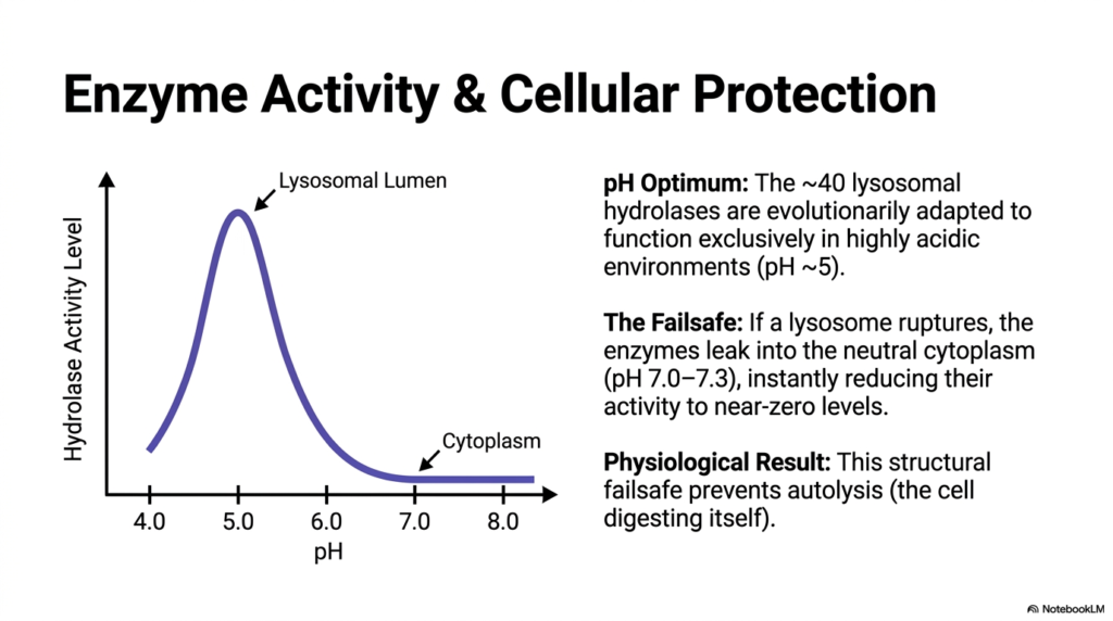
This slide visually graphs the relationship between pH levels and the activity of the enzymes housed within Lysosomes. The bell-shaped curve clearly demonstrates an evolutionary adaptation: these roughly forty acid hydrolases have a distinct pH optimum around 5.0. This means their catalytic efficiency peaks exclusively in the highly acidic environment within the lysosomal lumen. As the surrounding environment approaches the neutral pH of the cytoplasm, the biological activity of these enzymes drops dramatically, almost reaching zero.
The strict pH dependency of these enzymes provides an ingenious structural failsafe for the cell. If the membrane of a Lysosome were to tear or rupture, releasing its destructive payload, disaster might seem imminent. However, because the surrounding cytoplasm sits at a neutral pH of 7.0 to 7.3, the leaked hydrolases instantly denature or lose their functional shape. This immediate loss of enzyme activity serves as an automatic off-switch, neutralizing a potentially lethal threat to the cellular machinery.
The physiological result of this evolutionary design is the prevention of autolysis, a fatal process in which a cell essentially digests itself from the inside out. By locking the optimal functionality of these enzymes to a specific, artificially maintained acidic range, the cell ensures that degradation only happens within the safe confines of Lysosomes. This biochemical adaptation highlights the precision of cellular engineering, where chemical limits are used just as effectively as physical barriers to control potent reactions.
For medical students, understanding this pH-dependent failsafe is critical when studying cellular injury and necrosis. While a single ruptured vesicle is easily handled by this pH buffer system, massive cellular trauma can overwhelm the cytoplasm, leading to a catastrophic drop in overall cellular pH. When the entire cell becomes acidic, the lysosomal failsafe fails, and widespread autolysis ensues. Recognizing this delicate balance of hydrogen ion concentration is key to understanding both normal physiology and cellular death.
Slide 5: The Three Stages of Enzymatic Degradation in Lysosomes
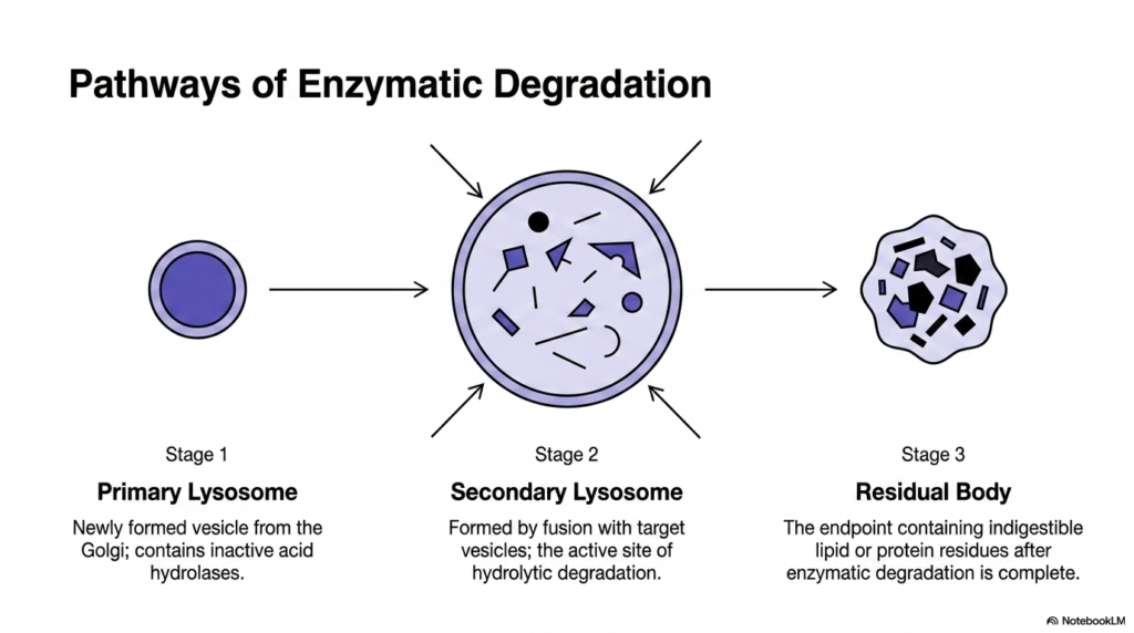
The biochemical journey of Lysosomes is dynamic, progressing through three distinct stages of maturation and degradation, as shown here. The cycle begins with Stage 1, the formation of the Primary Lysosome. This structure is a newly formed vesicle budding directly from the Golgi apparatus. At this initial phase, the vesicle contains a full complement of acid hydrolases, but these enzymes remain entirely inactive. The primary structure is essentially a biological delivery vehicle, primed and waiting to dismantle a specific target.
The critical transformation occurs during Stage 2, creating the Secondary Lysosome. This structure forms when the primary vesicle physically fuses with a target vesicle carrying material destined for destruction. It is within these Secondary Lysosomes that the active site of hydrolytic degradation is established. The internal pH drops, the enzymes activate, and the complex process of breaking down macromolecules into fundamental biological building blocks begins in earnest. This stage represents the functional peak of the organelle’s lifecycle.
Eventually, the digestive process reaches its natural conclusion at Stage 3, resulting in a Residual Body. Even the powerful enzymes within Lysosomes cannot break down every possible substance. The residual body is the final endpoint, containing indigestible lipid or protein residues left after active enzymatic degradation is complete. These remnants represent the absolute limits of the cellular digestive capacity and must be managed by the cell either through storage or eventual excretion.
Understanding this three-stage pathway highlights that Lysosomes are not static organelles but rather transient functional states. The transition from an inactive primary carrier to an actively digesting secondary structure, and finally to an exhausted residual body, demonstrates continuous metabolic flow. Recognizing this morphological evolution is essential for students tracking how waste is systematically isolated, aggressively dismantled, and ultimately packaged away by the intricate cellular waste management system.
Slide 6: Autophagy and Heterophagy Handled by Lysosomes
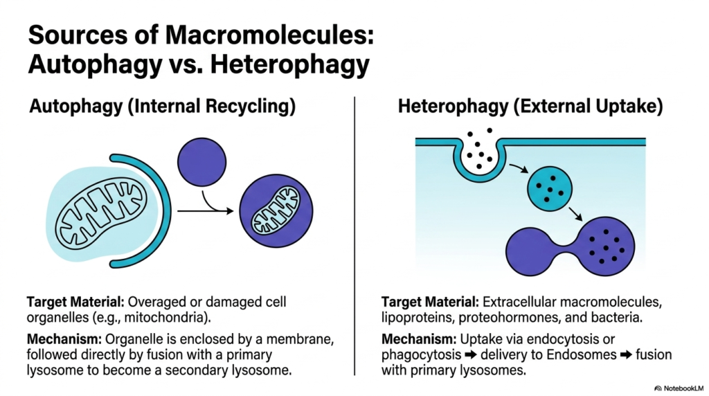
Cells must constantly process biological material originating from both internal and external sources, and Lysosomes sit firmly at the metabolic center of both major pathways. This slide clearly contrasts Autophagy, the necessary process of internal recycling, with Heterophagy, the systemic processing of external uptake. During autophagy, the cell targets its own overaged or severely damaged organelles, such as failing mitochondria. This constant internal housekeeping is vital for overall cellular health, ensuring that defective machinery does not accumulate and cause massive metabolic distress.
The mechanism of autophagy involves enclosing the targeted cellular structure within a double membrane to isolate it from the cytoplasm. This isolated package then fuses directly with primary Lysosomes. The resulting fusion creates a highly active secondary structure where the organelle is dismantled. By constantly turning over its internal components, the cell uses lysosomes to scavenge nutrients during starvation and to maintain peak functional efficiency during normal metabolic operations.
Conversely, heterophagy deals with materials brought in from the extracellular environment. The cell membrane continuously captures external macromolecules, lipoproteins, proteohormones, and even whole bacteria through processes known as endocytosis or phagocytosis. These external targets are encapsulated into endosomes, which subsequently travel deep into the cytoplasm to fuse with waiting Lysosomes. This external uptake pathway serves dual purposes: harvesting nutrients from the extracellular fluid and destroying invasive pathogens that threaten cellular integrity.
For students studying immunology and cellular metabolism, mastering the distinction between these two intersecting pathways is absolutely essential. Whether the human cell is digesting its own exhausted mitochondria to survive periods of intense starvation or actively engulfing dangerous bacterial invaders to protect the broader organism, Lysosomes serve as the universal biological executioners. Recognizing how these adaptable organelles seamlessly handle completely diverse biological materials underscores their indispensable, dual role in both cellular nutrition and innate immune defense.
Slide 7: Lysosomes Biogenesis Step 1 Synthesis in the rER
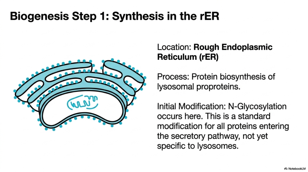
Building the destructive payload of Lysosomes requires precision engineering, beginning at the Rough Endoplasmic Reticulum (rER). This slide illustrates the very first step in biogenesis, focusing on the initial synthesis of digestive enzymes. The rER serves as the primary manufacturing hub for the basic protein chains of lysosomal proproteins. Ribosomes studded along the endoplasmic reticulum membrane translate messenger RNA into the raw, unfolded protein sequences that will eventually become potent hydrolases.
At this early stage, the proteins destined for Lysosomes are completely inactive and simply known as proproteins. Once synthesized, they undergo their first chemical modification within the lumen of the rER: N-glycosylation. During this process, complex sugar trees are attached to specific nitrogen atoms on the protein chain. This addition of carbohydrates helps fold the newly minted enzyme into a stable, three-dimensional structure necessary for its future journey through the cell.
It is crucial for students to recognize that N-Glycosylation is a standard biological modification for nearly all proteins entering the secretory pathway. At this specific point in the rER, the newly created proproteins lack any unique identification that designates them specifically for Lysosomes. They are structurally indistinguishable from normal proteins destined for the cell membrane or extracellular secretion. The true biochemical sorting that defines their final destination has not yet begun.
This initial synthesis phase strongly highlights the shared biochemical origins of all secretory and organelle-bound proteins. The Rough Endoplasmic Reticulum acts as a massive, general assembly line, rapidly producing an incredibly wide array of functional protein products. The specialized enzymes that will eventually arm the mature Lysosomes start their cellular lives completely anonymously among tens of thousands of other standard cellular proteins. Understanding this universal starting point sets the perfect stage for appreciating the highly specific, subsequent chemical tagging required to route them correctly.
Slide 8: The cis-Golgi Target in the Biogenesis of Lysosomes
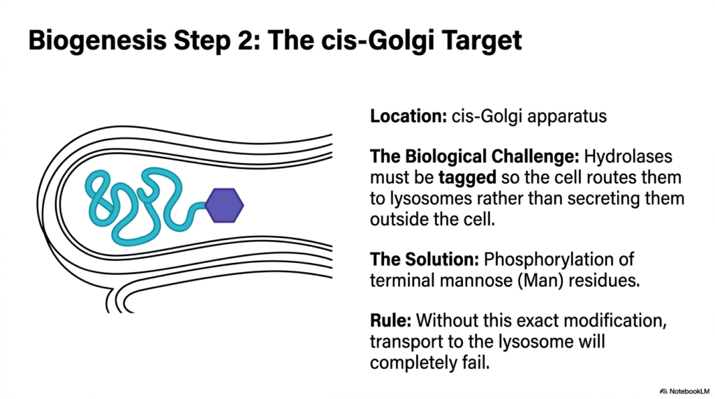
After leaving the Endoplasmic Reticulum, the newly synthesized proproteins arrive at the cis-Golgi apparatus, the critical sorting center for Lysosomes. This slide outlines the primary biological challenge the cell faces at this junction. Thousands of different proteins pass through the Golgi, and the cell must actively route the dangerous hydrolases away from the standard secretory pathway. If the cell fails to intercept these specific enzymes, they will be mistakenly secreted entirely outside the cell, leaving the cell defenseless.
To solve this massive logistical challenge, the cis-Golgi employs a highly specific biochemical tagging system solely for proteins destined for Lysosomes. The solution relies on the precise phosphorylation of terminal mannose residues attached to the enzyme. Earlier in the rER, simple sugar chains containing mannose were added to the protein. Now, inside the cis-Golgi, specialized enzymes recognize the unique shape of the lysosomal hydrolases and attach a phosphate group specifically to those mannose sugars.
This specific chemical addition creates the critical Mannose 6-phosphate tag. The rule of this biological sorting system is absolute and unforgiving. Without this exact structural modification, transport to the Lysosomes will completely fail. The enzymes would simply follow the default pathway out of the cell, leading to severe metabolic consequences. The phosphorylation event serves as an unforgeable molecular barcode, indicating that the protein is meant for internal destruction.
For students studying advanced biochemistry, the rapid events unfolding in the cis-Golgi perfectly illustrate the absolute elegance of molecular recognition. The cell utilizes a remarkably simple chemical modification—the strategic addition of a single phosphate group—to permanently alter the final destiny of massive, complex protein structures. This rigorous tagging mechanism ensures that the devastating power of digestive enzymes is safely corralled and precisely directed toward developing Lysosomes, showcasing the remarkable fidelity and precision of eukaryotic intracellular sorting networks.
Slide 9: Creating the Tag for Lysosomes via Reaction 1
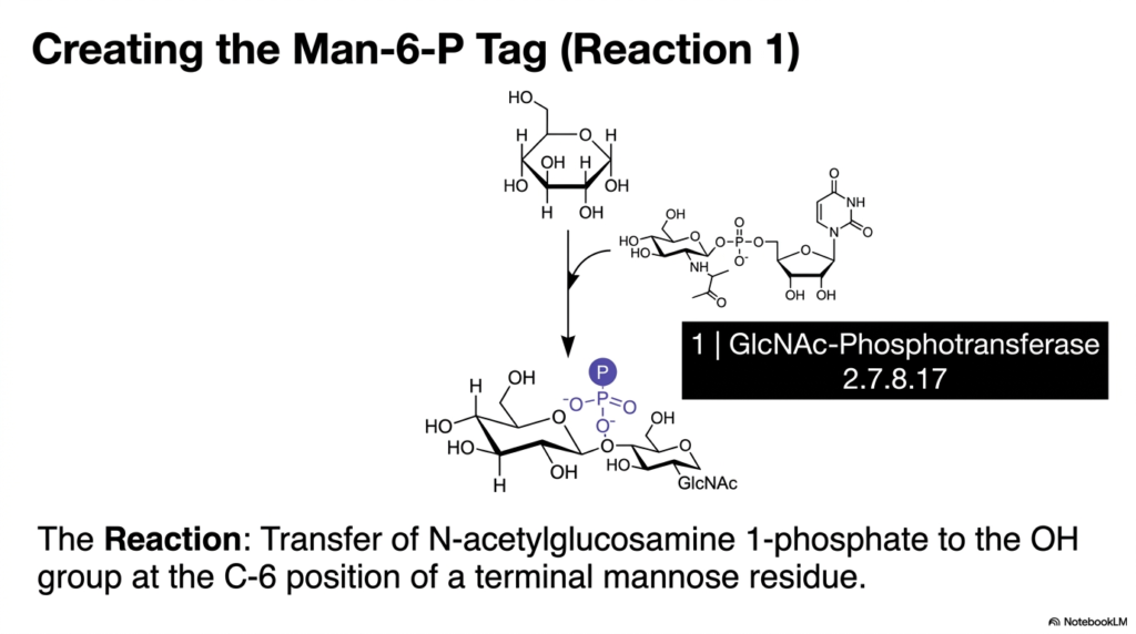
The molecular tagging of enzymes destined for Lysosomes is a complex two-step process, with this slide detailing the crucial first biochemical reaction. The visual highlights the specific molecular structures involved in creating the vital sorting signal. The primary actor in this first step is an enzyme known as GlcNAc-Phosphotransferase, officially classified under the enzyme commission number 2.7.8.17. This specific transferase initiates the formation of the essential chemical barcode required for accurate intracellular routing.
The biochemical reaction involves the precise transfer of an entire N-acetylglucosamine 1-phosphate molecule onto the existing sugar tree of the proprotein. Specifically, this bulky chemical group is attached to the hydroxyl (OH) group located exactly at the Carbon-6 position of a terminal mannose residue. This is not a random chemical event; the transferase specifically recognizes the unique three-dimensional conformational patch found only on the surface of enzymes intended for Lysosomes, ensuring absolute specificity.
At the end of Reaction 1, the protein destined for Lysosomes carries a somewhat masked signal. The phosphate group is now successfully attached to the mannose sugar, but it remains covered by the large N-acetylglucosamine moiety. This intermediate structure acts as a temporary protective cap during the ongoing chemical synthesis. Students must recognize that while the phosphate is present, the sorting signal is not yet fully active or recognizable by the cellular transport machinery.
Understanding this initial transferase reaction is exceptionally critical for medical students because it represents the primary molecular bottleneck in the successful biogenesis of Lysosomes. If the specific GlcNAc-Phosphotransferase enzyme is defective due to an inherited genetic mutation, the entire tagging process halts immediately. The cell totally loses its ability to mark any of its newly minted hydrolases, leading to a catastrophic, systemic failure in spatial sorting and a complete lack of functional digestive organelles, profoundly disrupting basic cellular survival.
Slide 10: Finalizing the Tag for Lysosomes via Reaction 2
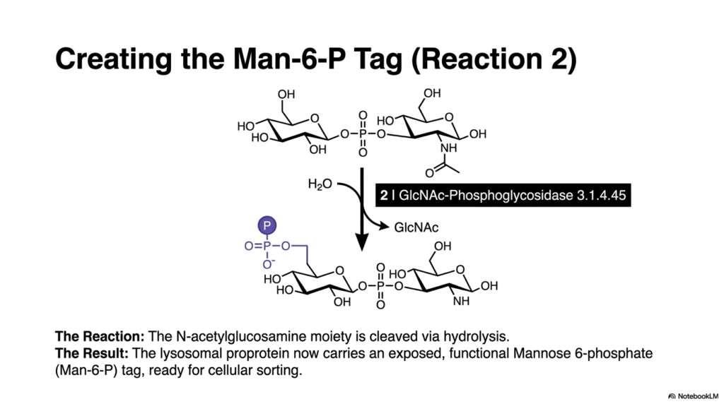
The intricate biochemical tagging process for Lysosomes concludes with the second critical reaction outlined in this slide. Following the initial transfer, the developing proprotein carries a masked signal that must be unveiled. This crucial unmasking is performed by a secondary enzyme, GlcNAc-Phosphoglycosidase, which is classified as 3.1.4.45. The sole purpose of this highly specific glycosidase is to finalize the molecular barcode, ensuring the digestive hydrolases can be accurately detected by the cellular transport network.
The chemical mechanism shown here is a straightforward hydrolysis reaction. The GlcNAc-Phosphoglycosidase enzyme actively cleaves the large N-acetylglucosamine moiety away from the underlying structure. The addition of a water molecule aggressively breaks the chemical bond, releasing the bulky GlcNAc group into the surrounding Golgi lumen. Stripping away this temporary protective cap permanently exposes the critical phosphate group attached directly to the sixth carbon of the mannose sugar.
The ultimate result of this two-step enzymatic process is the creation of a fully functional Mannose 6-phosphate (Man-6-P) tag. With the phosphate group now completely exposed and properly positioned, the lysosomal proprotein is officially ready for cellular sorting. This completed molecular identifier is the mandatory passport required for entry into the Lysosomes. Without this exposed phosphate, the protein would remain invisible to the specialized receptor networks waiting further along the Golgi apparatus.
For students analyzing deep cellular pathways, this elegant two-step mechanism perfectly illustrates the extreme biological caution the cell exercises when handling the dangerous enzymes of Lysosomes. By actively requiring two entirely separate, highly specific enzymes to successfully create the final sorting signal, the cell significantly reduces the risk of accidentally tagging random, vital structural proteins for destruction. This complex biochemical choreography guarantees that only the correct hydrolases receive the Man-6-P passport, maintaining strict, uncompromising control over the cellular digestive arsenal.
Slide 11: Receptor Sorting of Lysosomes in the trans-Golgi
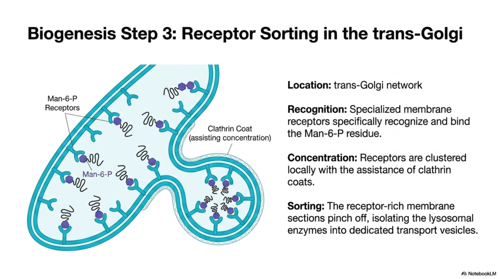
Once the biochemical tag is finalized, the sorting of Lysosomes shifts to the trans-Golgi network, as illustrated in this slide. This network serves as the cell’s final shipping department, where molecules are physically segregated into distinct outgoing vesicles. The location is critical because it is here that the newly exposed Mannose 6-phosphate (Man-6-P) tags are finally used. The environment within the trans-Golgi is specifically tailored to facilitate strong binding between the tagged enzymes and the cellular transport machinery.
The actual recognition process relies on specialized membrane proteins known as Man-6-P receptors. These receptors possess uniquely shaped binding pockets that specifically recognize and lock onto the exposed Man-6-P residues on the floating proproteins. As these receptors successfully capture the dangerous hydrolases, they begin to cluster together locally within the trans-Golgi membrane. This clustering ensures that enzymes intended for the Lysosomes are highly concentrated in a specific region of the membrane, preventing their accidental secretion.
This concentration of receptors is physically assisted by the formation of clathrin coats on the cytosolic side of the Golgi membrane. The clathrin proteins assemble into a rigid, cage-like structure that mechanically pulls the flat lipid bilayer into a distinct, curved bud. As this receptor-rich section of the membrane bulges outward, it effectively traps the lysosomal proproteins inside. The structural support from the clathrin coat is absolutely essential for gathering the widespread receptors into a tightly organized delivery package.
Finally, the sorting process concludes when these receptor-rich membrane sections physically pinch off from the main trans-Golgi body. This crucial scission event permanently isolates the potent lysosomal enzymes into dedicated, independent transport vesicles. The hydrolases are now safely packaged, securely bound to their receptors, and actively traversing the cytoplasm on a direct trajectory toward the developing Lysosomes. This dynamic vesicle formation marks the successful transition from chemical tagging to physical cellular transport.
Slide 12: Transport and Dissociation to the Endolysosome
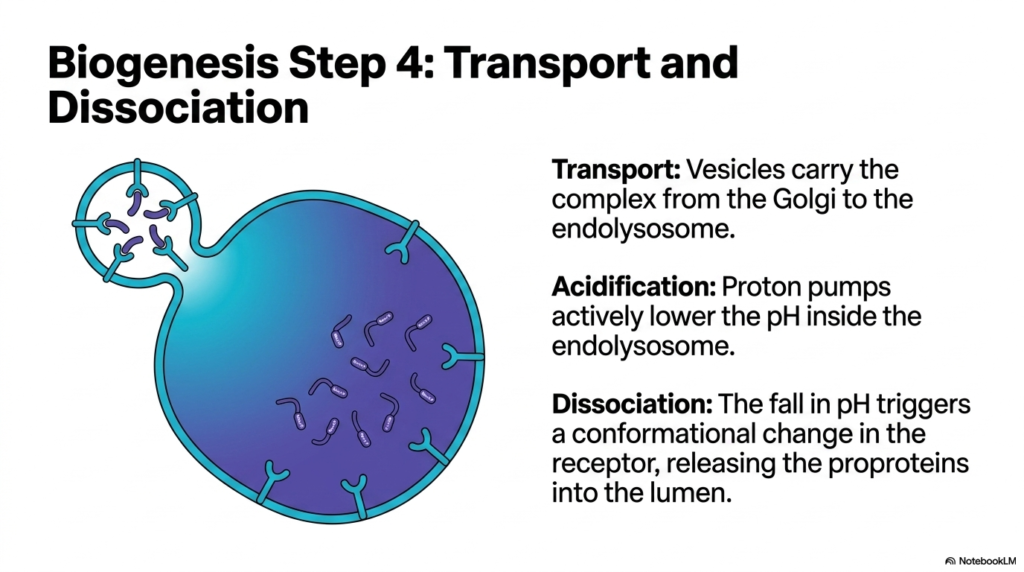
The physical transport of digestive enzymes to mature Lysosomes involves a crucial intermediary stage known as the endolysosome, as shown in this slide. After pinching off from the trans-Golgi network, the dedicated transport vesicles carry the tightly bound receptor-proprotein complexes through the cytoplasm. These small vesicles quickly locate and fuse with larger, waiting structures to form the endolysosome. This fusion event physically delivers the dangerous hydrolases to the proper cellular compartment, but the enzymes remain stubbornly bound to their transport receptors.
To release the enzymes and complete delivery to Lysosomes, the cell rapidly acidifies the new compartment. ATP-driven proton pumps, similar to those discussed in earlier slides, actively flood the endolysosome lumen with hydrogen ions, aggressively lowering the internal pH. This targeted drop in pH is not just about creating a corrosive environment; it is a vital chemical trigger specifically designed to alter the physical shapes of the proteins involved in the transport process.
The increasing acidity causes a profound structural dissociation event. As the pH drops, the highly sensitive Man-6-P receptors undergo a sudden, dramatic conformational change. Their physical shape shifts, forcing their specialized binding pockets to break apart and release the Mannose 6-phosphate tags. This acidic trigger successfully uncouples the receptor from the enzyme, permanently releasing the vital proproteins freely into the fluid lumen of the developing Lysosomes, effectively completing the physical delivery phase.
Understanding this strict pH-dependent dissociation is absolutely critical for medical students studying the intricate dynamics of intracellular transport. If the endolysosome fails to acidify properly due to metabolic stress, the receptors simply cannot release the bound hydrolases. The entire digestive payload would remain permanently locked to the inner membrane, rendering the future Lysosomes completely useless. The elegant use of rapidly rising acidity to mechanically force the transport receptors open ensures that the toxic enzymes are only uncoupled once safely sealed inside their final, heavily fortified biological destination.
Slide 13: Receptor Recycling and Maturation of Primary Lysosomes
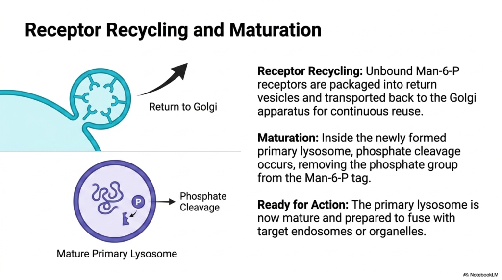
Efficiency is a hallmark of cellular biology, and the biogenesis of Lysosomes relies heavily on massive receptor recycling, as shown in this slide. After the acidic environment forces the dissociation of the enzymes, the empty Man-6-P receptors do not remain in the harsh lumen. Instead, the cell rapidly gathers these unbound receptors, packages them into small return vesicles, and actively transports them backward to the trans-Golgi apparatus. This constant retrograde transport ensures an endless supply of receptors for continuous protein sorting.
While the receptors return to the Golgi, the newly formed primary Lysosomes undergo a final, vital maturation phase. Within the acidic lumen, the free-floating proproteins undergo targeted phosphate cleavage. Specialized resident acid phosphatases systematically strip the phosphate groups entirely away from the Mannose 6-phosphate tags. This removal is crucial because it permanently traps the enzymes inside the vesicle; without the tag, they cannot accidentally bind to any stray receptors and escape back to the Golgi network.
The cleavage of the phosphate group marks the definitive transition into mature, fully armed primary Lysosomes. The previously masked and tightly regulated enzymes are now fully liberated within their highly acidic, optimal environment. The primary organelle is now fully mature structurally, packing a concentrated, potent arsenal of active acid hydrolases. It requires no further chemical modifications or sorting signals from the cell to perform its biological function; it is merely waiting for a suitable metabolic target.
Medical students must firmly grasp that this final biochemical step officially completes the remarkable cellular assembly line of Lysosomes. What initially began as a completely generic protein chain in the rough ER is now a highly specialized, extremely dangerous biological tool permanently locked inside a protected lipid vault. Fully ready for immediate action, this mature primary vesicle will patiently patrol the cytoplasm until it is specifically called upon to directly fuse with targeted endosomes or failing organelles, rapidly initiating the massive chemical breakdown essential for sustaining human life.
Slide 14: Clinical Correlation: Lysosomal Storage Diseases
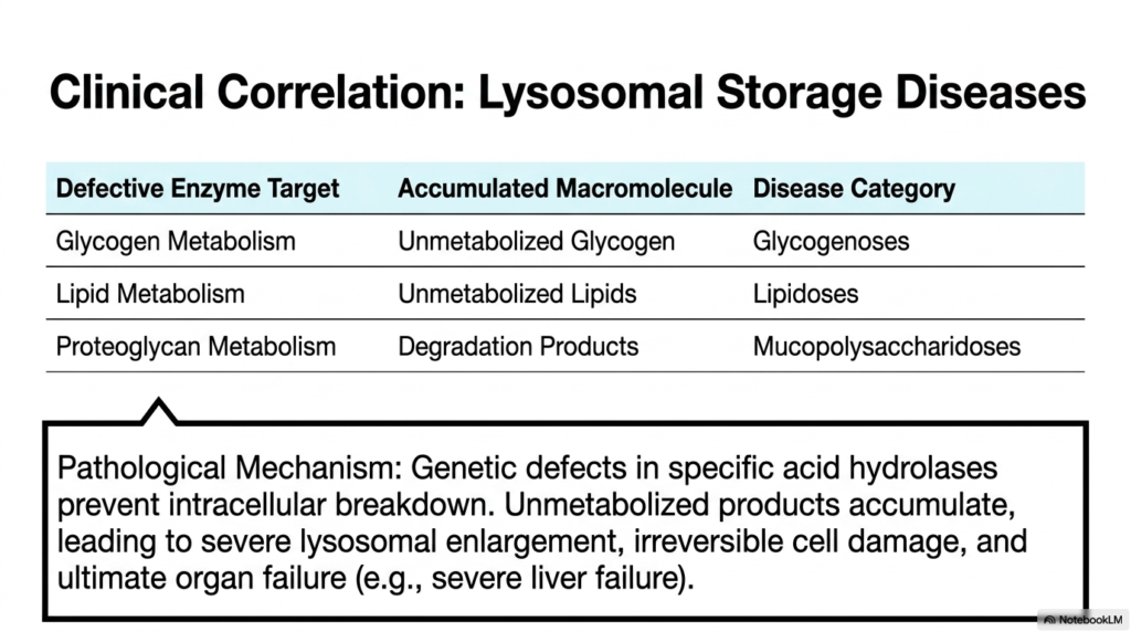
The profound importance of Lysosomes is most glaringly obvious when their mechanisms fail, leading to devastating clinical conditions known as Lysosomal Storage Diseases (LSDs). This slide details the direct pathological consequences of genetic defects within these critical organelles. When the cell harbors a mutation affecting even one specific acid hydrolase, the entire digestive pipeline stalls. The biological macromolecules that this missing enzyme would normally degrade begin to accumulate relentlessly inside the cell, completely overwhelming the organelle’s physical capacity.
The table highlights three primary disease categories directly linked to defective Lysosomes. Glycogenoses occur when defective enzymes fail to metabolize complex glycogen chains. Lipidoses arise from defects in lipid metabolism, causing toxic fatty substances to accumulate within neural tissues. Finally, Mucopolysaccharidoses result from an inability to break down complex proteoglycans found in connective tissues. Each category underscores how a single missing chemical tool within these tiny cellular stomachs can trigger systemic, organism-wide metabolic collapse.
The underlying pathological mechanism is heartbreakingly universal across virtually all identified Lysosomal Storage Diseases. As completely unmetabolized products rapidly accumulate, they directly cause severe and progressive physical enlargement of the internal Lysosomes. These bloated organelles massively balloon outward, mechanically crushing the surrounding cytoplasm and completely disrupting the normal, healthy architecture of the cell. This intense physical overcrowding leads to immense, unmanageable cellular stress, causing irreversible structural damage and ultimately triggering widespread, chaotic cell death across multiple vital organ systems, entirely destroying the patient’s biological health.
For aspiring physicians, grasping the link between cellular biochemistry and macroscopic symptoms is absolutely vital. Patients suffering from these genetic defects in Lysosomes frequently present with tragic, cascading clinical symptoms, culminating in severe organ failure, particularly overwhelming liver and brain failure. By understanding the exact biochemical breakdowns occurring at the microscopic level, medical students can better comprehend the relentless progression and immense clinical severity of these inherited metabolic disorders.
Slide 15: Synthesis of the Lysosomal Lifecycle
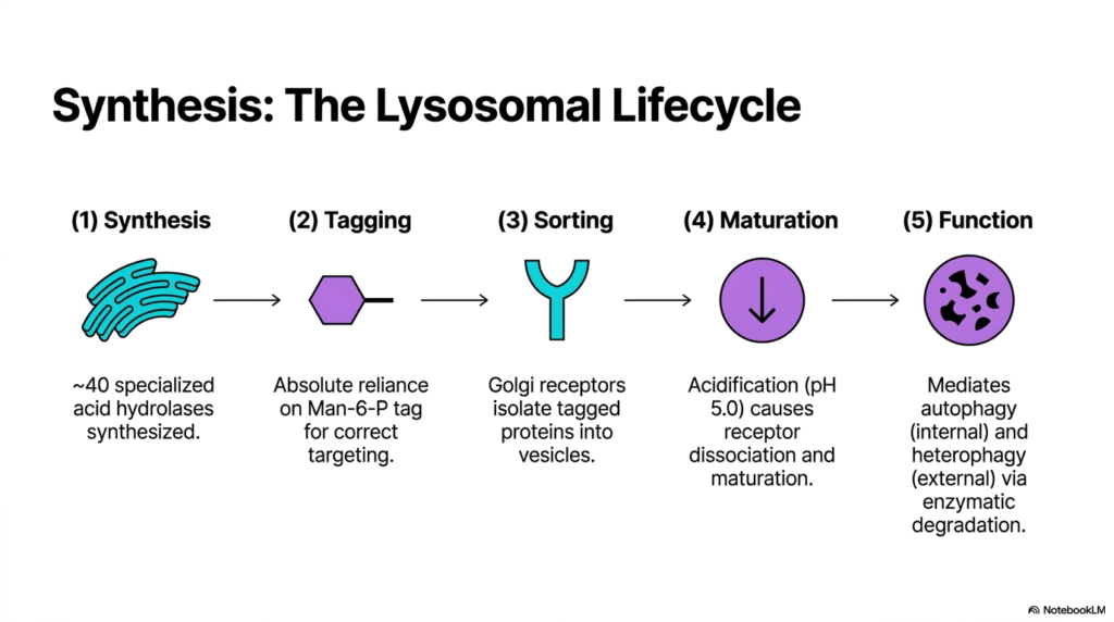
This concluding slide masterfully synthesizes the entire complex lifecycle of Lysosomes, summarizing the five essential steps required for their biological function. The journey begins with Synthesis in the endoplasmic reticulum, where roughly forty highly specialized acid hydrolases are carefully manufactured. This initial step represents the immense metabolic investment the cell makes to guarantee it possesses a comprehensive suite of destructive tools capable of dismantling any biological polymer it encounters throughout its life.
The second and third steps emphasize the absolute necessity of precision routing to build Lysosomes. Tagging involves the rigorous addition of the Mannose 6-phosphate signal, an absolute prerequisite for correct cellular targeting. Without this tag, the entire system collapses. Following tagging is the Sorting phase, where specialized Golgi receptors specifically identify the Man-6-P tags and actively isolate these dangerous proteins into highly secure, dedicated transport vesicles, safely shielding the rest of the cytoplasm from accidental digestion.
The lifecycle concludes with Maturation and Function, firmly establishing the biological roles of Lysosomes. Maturation is driven by a massive influx of protons, lowering the internal pH to 5.0. This extreme acidification chemically forces receptor dissociation and fully arms the waiting hydrolases. Finally, the mature organelles execute their Function: mediating both internal autophagy to recycle damaged cellular components and external heterophagy to digest engulfed nutrients and destroy invading pathogens through relentless enzymatic degradation.
Reviewing this beautiful, entire biological sequence provides medical students with a profoundly holistic understanding of eukaryotic cellular waste management. The continuous lifecycle of Lysosomes is a dynamic, highly regulated biochemical assembly line that perfectly blends complex molecular genetics, intricate chemical tagging, rapid active membrane transport, and incredibly robust structural failsafes. Mastering these deeply interconnected five steps allows future biological scientists and practicing doctors to truly appreciate how the microscopic world of the cell expertly balances potent, incredibly dangerous destructive capabilities with vital self-preservation to gracefully sustain human life.
Please read our Content Disclaimer Statement.
Check out our social media channels: