159. Oncogenes: Molecular Drivers of Cellular Transformation
Cancer often feels like a mysterious breakdown of the body, but at its core lies a logical failure in cellular communication. Why do healthy, well-behaved cells suddenly transform into aggressive tumors that divide without stopping? The answer usually points to altered signaling components within our genome. In this slide deck, we will map out how normal cellular growth controls fail and explore the key biochemical pathways driven by oncogenes.
Slide 01: Oncogenes: Molecular Drivers of Cellular Transformation
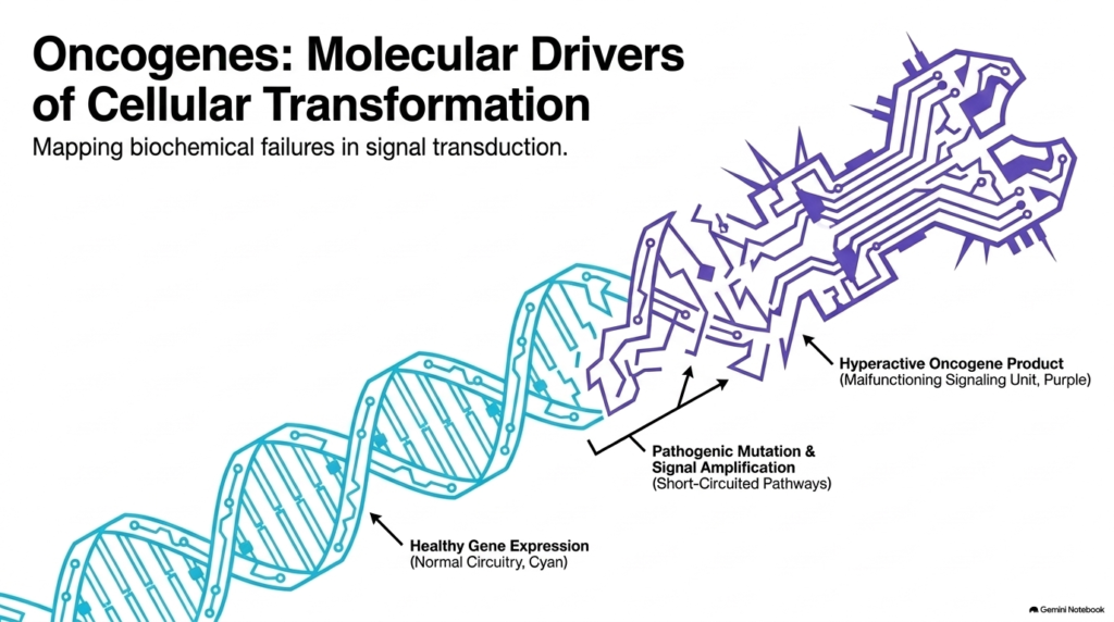
When a normal cell receives a signal to divide, it relies on an intricate network of genes that act like clean electrical wiring. Healthy gene expression keeps growth signals balanced and properly regulated. However, pathogenic mutations can short-circuit this circuitry, turning friendly genetic instructions into disease-driving signals. These mutated, hyperactive genetic variants are broadly classified as oncogenes.
Understanding how oncogenes disrupt cellular stability is essential to unraveling cancer’s underlying biochemistry. In healthy tissues, signaling proteins switch on only when needed and quickly shut off after completing their task. When genes undergo structural mutations or signal amplification, they produce malfunctioning signaling units that remain permanently turned on. This hyperactive state floods the cell with continuous growth signals, bypassing the regulatory checkpoints that maintain tissue balance.
As these short-circuited pathways multiply, the affected cell begins its transition toward malignant cellular transformation. Activated oncogenes act as persistent genetic accelerators, pushing the cell to copy its genome and divide repeatedly without waiting for external growth factors. Over time, this constant signal amplification overrides the protective mechanisms that keep tissue growth under control. By studying how oncogenes hijack normal signaling networks, biochemists can identify specific molecular targets to disarm these rogue proteins.
Slide 02: Proto-oncogenes vs. Oncogenes in Cellular Regulation
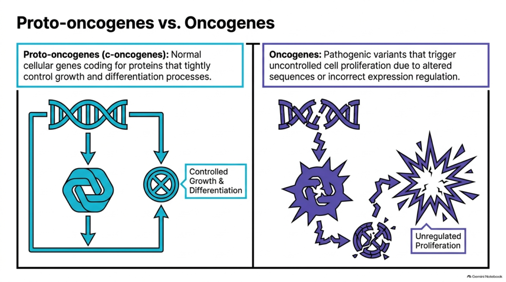
To understand how cancer develops, we must first distinguish between healthy genes and their disease-causing counterparts. Normal cells rely on proto-oncogenes, also called c-oncogenes, which encode vital proteins that regulate cell growth, division, and maturation. These proteins function like carefully calibrated switches, ensuring tissue growth occurs only when needed for physiological development or tissue repair.
Problems arise when environmental toxins, radiation, or DNA replication errors alter the genetic sequence of these essential genes. When a proto-oncogene gains a gain-of-function mutation or loses normal regulatory control, it becomes a dangerous variant. Oncogenes are mutated genes that drive unchecked cell division and proliferation. While healthy genes tightly control cellular growth, these hyperactive variants generate rogue signals that bypass normal safety mechanisms.
The fundamental difference lies in how these two gene states affect overall cellular behavior. Proto-oncogenes promote orderly tissue maintenance and respond promptly to inhibitory signals from neighboring cells. In contrast, oncogenes force the cell into an endless cycle of unregulated proliferation. The cell loses its ability to pause, repair damaged DNA, or undergo programmed cell death. This continuous, unchecked division lays the biochemical foundation for tumor development, transforming a once cooperative cell into a self-serving clone.
Slide 03: Mechanisms of Genetic Transformation into Oncogenes
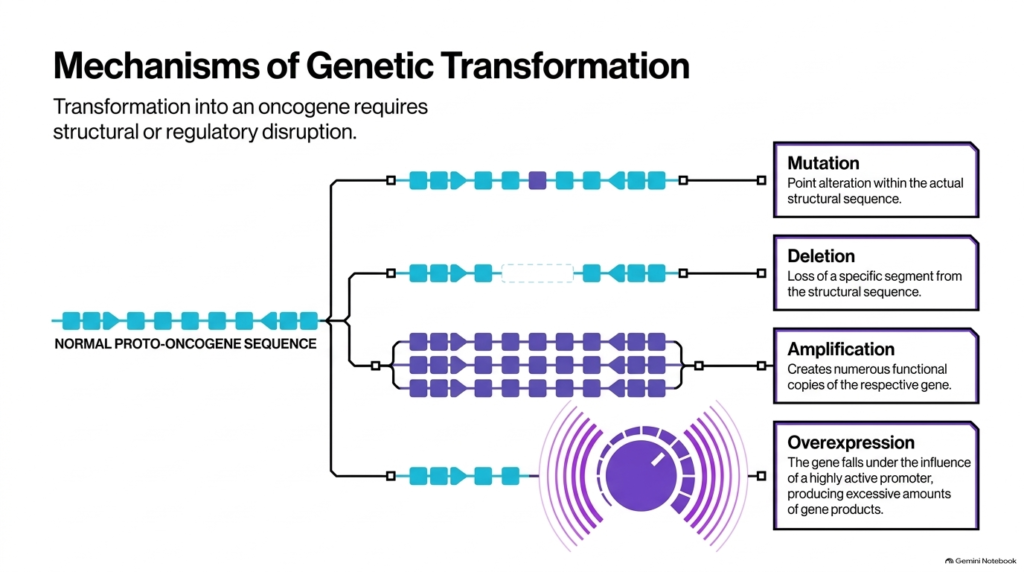
A normal cellular gene does not transform into a disease driver by chance; it requires specific structural or regulatory disruptions. Biochemists have identified four primary genetic mechanisms that convert normal cellular genes into hyperactive oncogenes. Each mechanism alters either the protein’s primary structure or the total amount of protein the cell produces.
The first mechanism is a point mutation, in which a single nucleotide substitution changes an amino acid in the protein sequence. This subtle change can lock the resulting protein into a permanently active conformation. The second mechanism involves deletions, where the loss of a specific gene segment removes critical inhibitory domains. Without these built-in regulatory controls, the protein signals without stopping. Both point mutations and deletions directly change how the protein functions inside the cell.
The remaining two mechanisms focus on producing excessive quantities of normal or altered proteins. Gene amplification occurs when chromosomal errors create numerous extra copies of a gene, leading to massive protein production. Overexpression happens when a gene falls under the influence of a highly active promoter, driving continuous transcription. Through these pathways, normal cellular genes become hyperactive oncogenes. As these mutated oncogenes accumulate, they overwhelm the delicate signaling networks that keep cellular division under strict control.
Slide 04: The Viral Origin and Nomenclature of Oncogenes
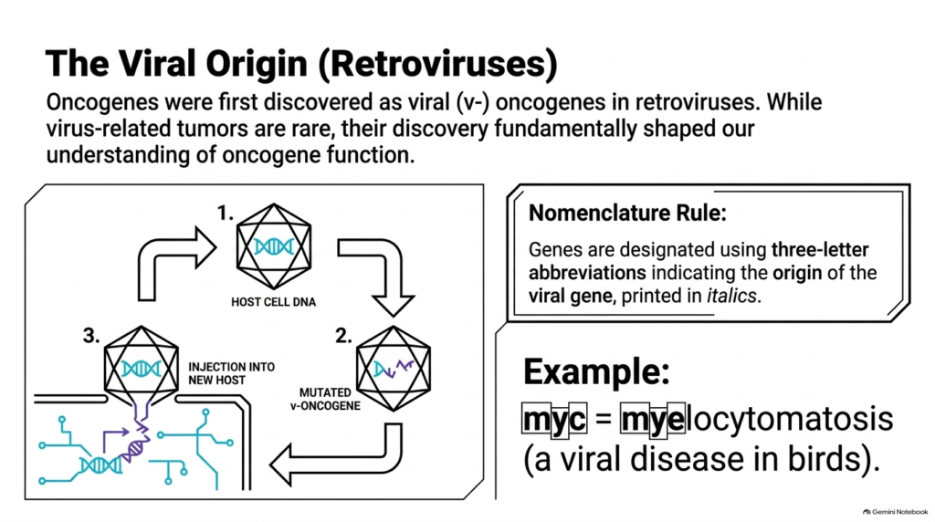
The discovery of these cancer-driving genes has a fascinating history rooted in virology. Researchers studying tumor-causing retroviruses found that viruses carry specific genetic sequences that can transform healthy host cells into cancer cells. Researchers named these viral sequences v-oncogenes, marking a historic breakthrough in modern cancer biology. Although virus-induced tumors are relatively rare in humans, studying retroviruses revealed how rogue genes disrupt normal cell behavior.
Further research demonstrated that retroviruses actually stole these signaling genes from host organisms during ancient infections. When a virus picks up a host proto-oncogene, it often mutates the sequence and reintroduces it as a hyperactive driver. To organize these discoveries, scientists developed a standardized naming rule. Scientists designate every gene using a three-letter italicized abbreviation based on the viral disease in which it was first isolated.
For example, the famous myc gene derives its name from avian myelocytomatosis, a viral disease affecting birds. Understanding this nomenclature helps researchers identify related gene families across different species. By comparing viral genes with cellular genes, biochemists realized that human cancers often rely on the same signaling pathways retroviruses manipulate. Thus, early viral research provided the fundamental framework for modern study into how oncogenes drive malignant transformation. Analyzing these ancient retroviral sequences revealed general principles of how cellular oncogenes subvert normal growth controls.
Slide 05: The “Two-Hit” Requirement for Transformation by Oncogenes
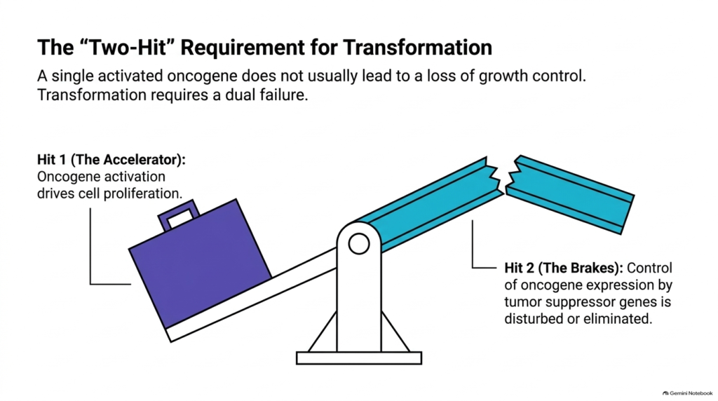
A single genetic mutation rarely transforms a healthy cell into a fully malignant tumor. Cells have robust, overlapping protective networks that monitor signaling and repair cellular damage. Consequently, cellular transformation typically requires a dual failure, often described as a two-hit model. Activated oncogenes serve as the first hit, providing the initial drive toward uncontrolled cell division.
Think of cell growth as a heavy vehicle moving along a highway. The first hit acts like a jammed accelerator pedal, constantly pushing the engine to run faster. Expressed oncogenes generate powerful growth signals that urge the cell to divide continuously. However, if the brakes are still functional, the cell can slow down, pause the cell cycle, or trigger self-destruction before damage spreads. The system only collapses when the biological emergency brakes fail.
The second hit occurs when tumor suppressor genes lose their ability to control or silence excessive growth signals. Tumor suppressors normally counteract hyperactive pathways by halting division or repairing mutations. When a cell suffers both an activated accelerator and broken brakes, the balance tilts irreversibly toward malignant transformation. Understanding how oncogenes interact with tumor suppressors explains why healthy cells usually resist single mutations and highlights why cancer therapies must target multiple signaling checkpoints simultaneously.
Slide 06: Architectural Overview of Signal Transduction by Oncogenes
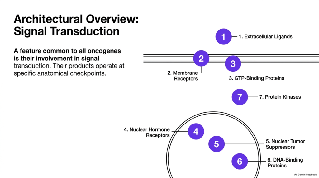
A universal feature unifying all cancer-causing genes is their direct involvement in signal transduction pathways. Signal transduction is the multi-step biochemical process by which a cell receives external cues, transmits them across the membrane, and converts them into precise genetic responses. The protein products of various oncogenes operate at specific anatomical checkpoints along this complex communication chain, disrupting normal regulation at every cellular level.
These signaling checkpoints span the entire architecture of the cell, starting outside the plasma membrane and reaching deep into the nucleus. Extracellular growth factors and transmembrane receptors receive initial signals, while cytosolic GTP-binding proteins and protein kinases relay the message through chemical modifications. Meanwhile, nuclear hormone receptors and DNA-binding transcription factors directly execute gene expression programs inside the cell nucleus, supported or opposed by nuclear tumor suppressors.
When mutations strike any of these checkpoints, the entire signaling cascade becomes compromised. A defect at a single node can send false signals down the entire chain, mimicking the presence of external growth factors. By classifying signaling components by location and biochemical activity, researchers can map exact failure points. This spatial overview shows that oncogenes are not random mutations but structural disruptions at key nodes in cellular communication networks. Recognizing these specific cellular locations helps biochemists categorize how different oncogenes drive uncontrolled proliferation.
Slide 07: Class 1 Extracellular Ligands as Oncogenes
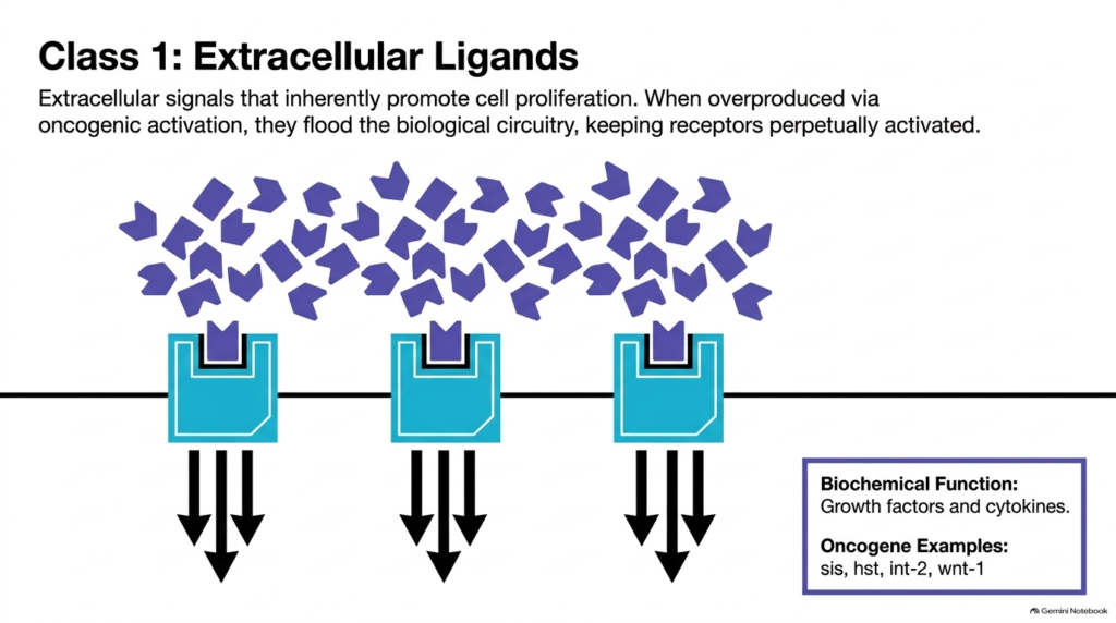
The first major class of cancer-driving proteins consists of overproduced extracellular ligands, such as growth factors and cytokines. Normally, cells produce and release signaling molecules in strictly controlled quantities only when neighboring tissues require growth or repair. However, when specific genes become pathologically activated, cells begin overproducing these potent ligands, flooding the surrounding microenvironment with continuous growth cues as hyperactive oncogenes disrupt cellular quiescence.
This excess of extracellular ligands creates a dangerous autocrine or paracrine signaling loop. Surrounding transmembrane receptors become perpetually bound by ligands, keeping downstream biochemical machinery constantly engaged. The cell tricks itself into believing that external conditions demand rapid growth, even when no physiological need exists. This continuous ligand stimulation short-circuits normal cellular quiescence, driving surrounding cells into endless rounds of division and fostering tumor growth.
Classic examples of genes encoding hyperactive growth factors include sis, hst, int-2, and wnt-1. For instance, the sis gene encodes a subunit of platelet-derived growth factor. When overexpressed, it continuously stimulates target cells to divide without restraint. By overproducing these primary messenger molecules, class 1 oncogenes initiate the entire signaling cascade at the cell surface, keeping internal pathways permanently locked in an active growth state. Targeting these ligand-encoding oncogenes prevents the initial activation of cell-surface receptors.
Slide 08: Class 2 Membrane Receptors Transformed into Oncogenes
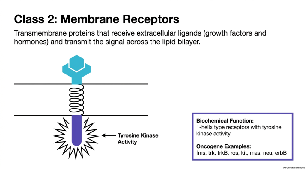
Transmembrane receptors act as vital gatekeepers, bridging the extracellular environment with the cell interior. Class 2 signaling components are single-helix transmembrane receptors with intrinsic enzymatic activity, specifically tyrosine kinase activity. In healthy tissues, these receptors remain dormant until an extracellular ligand binds to the outer domain, triggering receptor dimerization and activating the internal kinase domain.
When mutations alter the structural genes for these membrane receptors, the consequences for the cell are severe. Structural changes can cause receptor molecules to dimerize spontaneously or alter their internal kinase domains so they fire continuously without needing any ligand. Mutated membrane receptors act as potent oncogenes, sending relentless intracellular signals across the lipid bilayer even without external growth factors. When expressed in high numbers, these receptor-derived oncogenes lock downstream signaling networks in perpetual activation.
Biochemists have identified numerous gene examples within this class, including fms, trk, trkB, ros, kit, mas, neu, and erbB. For example, mutations in the neu gene produce a modified receptor that aggregates automatically, sending continuous growth commands to downstream targets. Because these membrane-bound kinases sit at the top of intracellular pathways, these hyperactive oncogenes amplify proliferation signals, making them prime targets for modern targeted cancer therapies.
Slide 09: Class 3 GTP-Binding Proteins as Oncogenes
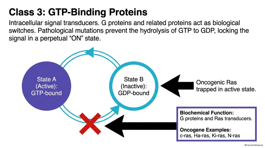
Inside the cell, signal transmission relies heavily on molecular switches that alternate between active and inactive states. Class 3 signal transducers are GTP-binding proteins, including the famous Ras protein family, which operate along the inner surface of the plasma membrane. Under normal conditions, these proteins bind guanosine triphosphate to become active, transmit a brief growth signal, and then hydrolyze GTP to guanosine diphosphate to turn off.
Pathological mutations in these molecular switches disrupt the essential GTPase activity needed to terminate signals. When point mutations strike critical codon positions, the mutated Ras protein loses its ability to hydrolyze GTP into GDP. As a result, the protein remains permanently trapped in its active, GTP-bound conformation. These altered transducers are among the most frequently mutated oncogenes in human clinical tumors.
The failure to shut down this molecular switch keeps downstream signaling cascades firing without interruption. Members of this gene family, such as c-ras, Ha-ras, Ki-ras, and N-ras, continuously stimulate downstream kinases that promote cell division and survival. Because trapped Ras proteins ignore normal shut-off signals, these oncogenes act as persistent internal engines of cell proliferation, making them a central focus of biochemical research and drug development. Inhibiting these GTPase-deficient oncogenes remains a top priority in molecular oncology.
Slide 10: Class 7 Protein Kinases as Intracellular Oncogenes
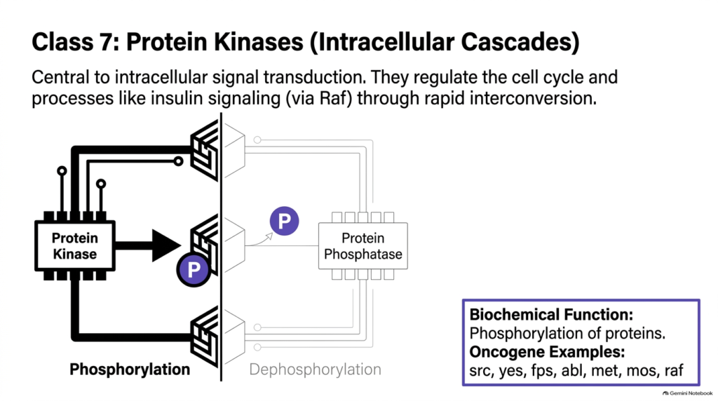
Once a growth signal crosses the cell membrane, protein kinases amplify it through intracellular phosphorylation cascades. Class 7 signaling components are cytosolic enzymes that transfer phosphate groups from ATP to specific amino acid residues on target proteins. This rapid enzymatic interconversion regulates crucial cell cycle decisions and metabolic pathways, such as insulin signaling mediated through the Raf protein pathway.
In normal cells, protein phosphatases quickly remove phosphate groups to reverse phosphorylation and restore baseline conditions. However, genetic mutations that alter kinase structural domains or regulatory regions can make these enzymes constitutively active. Hyperactive cytosolic kinases function as powerful oncogenes, continually phosphorylating target substrates regardless of upstream signals. This uncontrolled enzymatic activity overrides normal cellular checks, keeping metabolic pathways permanently primed for growth.
Key examples of cytosolic protein kinase genes include src, yes, fps, abl, met, mos, and raf. For instance, the src gene product was one of the earliest discovered tyrosine kinases capable of transforming cellular architecture when hyperactivated. When these intracellular cascades run unchecked, they flood the cytoplasm with active phosphorylated proteins. Understanding how these kinase-derived oncogenes operate has led to breakthrough targeted inhibitor drugs that block phosphate transfer and halt cancer growth. Disarming these hyperactive kinase oncogenes effectively cuts off internal relay signals.
Slide 11: Class 4 Nuclear Hormone Receptors as Oncogenes
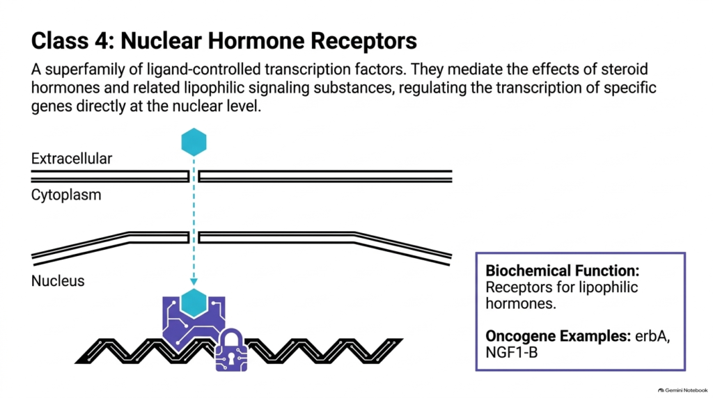
While many signaling pathways rely on membrane-bound receptors, lipophilic signaling molecules can diffuse directly through cellular membranes. Class 4 signaling components belong to a superfamily of nuclear hormone receptors that function as ligand-controlled transcription factors. These specialized receptors bind steroid hormones and other lipid-soluble substances, then migrate to the nucleus to bind DNA directly and regulate specific gene expression programs.
When structural alterations strike nuclear hormone receptor genes, their regulatory mechanisms break down dramatically. Mutated receptors may bind DNA tightly without requiring ligand binding, or they may permanently block normal developmental pathways by acting as dominant negative factors. These altered nuclear receptors act as oncogenes, directly disrupting transcription in the nucleus and promoting aberrant cell survival and proliferation. Expressed nuclear receptor oncogenes disrupt normal tissue maturation by hijacking transcriptional machinery.
Prominent gene examples in this category include erbA and NGF1-B. The erbA gene, originally identified in avian erythroblastosis virus, encodes a mutated thyroid hormone receptor. Unlike its normal cellular counterpart, the truncated ErbA protein cannot properly activate differentiation genes, trapping precursor cells in an immature, rapidly dividing state. By directly altering nuclear gene expression, these transcriptionally active oncogenes bypass cytoplasmic signaling cascades entirely to drive malignant growth.
Slide 12: Class 6 DNA-Binding Proteins as Nuclear Oncogenes
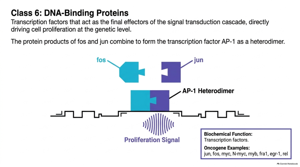
Every growth signaling cascade ultimately reaches the nucleus, where transcription factors directly control gene expression. Class 6 signaling components are DNA-binding proteins that act as the ultimate nuclear effectors of cell proliferation. Under healthy conditions, the cell rapidly synthesizes these nuclear factors in response to upstream growth signals and degrades them quickly once transcription is complete.
When mutations cause these nuclear proteins to be produced continuously or resist degradation, the cell experiences relentless transcriptional drive. Overexpressed nuclear factors function as potent oncogenes, binding directly to specific promoter sequences on target DNA to initiate cell division. A classic biochemical example is the AP-1 transcription factor complex, formed when the protein products of fos and jun combine into a functional heterodimer that binds DNA.
In addition to fos and jun, this critical class includes famous genes such as myc, N-myc, myb, fra1, egr-1, and rel. For example, the myc gene product regulates hundreds of genes involved in cell growth, metabolism, and protein synthesis. When these master transcriptional regulators escape normal expression controls, these nuclear oncogenes turn on the cell’s entire proliferative program, driving unchecked replication at the fundamental genetic level. Blocking these nuclear oncogenes remains a vital strategy for halting late-stage transcriptional driver events.
Slide 13: Class 5 Nuclear Tumor Suppressors and Anti-Oncogenes
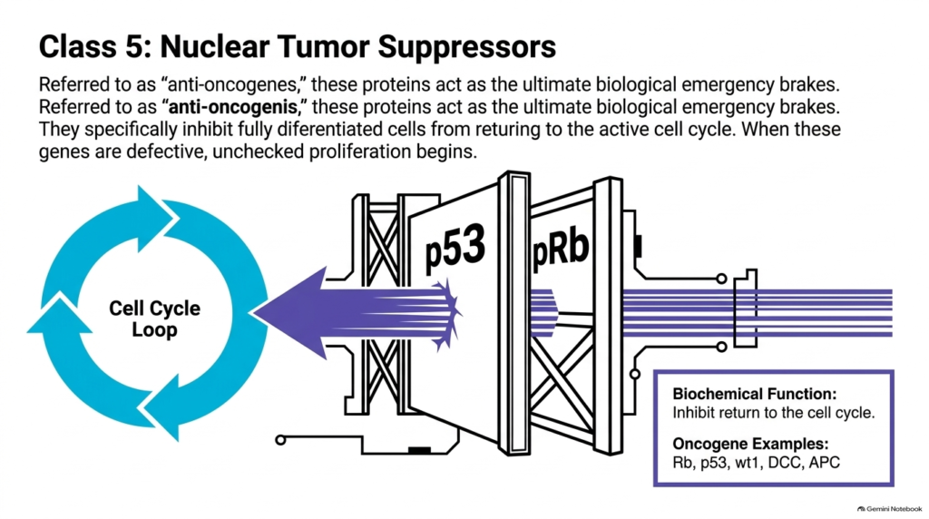
To counteract powerful pro-growth signals, cells rely on nuclear tumor suppressors, historically called anti-oncogenes. These nuclear proteins act as biological emergency brakes, preventing differentiated cells from inappropriately re-entering the cell cycle. Key proteins like p53 and the retinoblastoma protein, pRb, monitor genomic integrity and halt division or trigger cell death when they detect significant DNA damage.
When genetic mutations or viral interactions render these protective nuclear proteins defective, the cell loses its primary defense against hyperactive growth pathways. Without these emergency brakes, cells with damaged DNA can pass cell-cycle checkpoints without pausing for repair. While traditional growth drivers actively promote division, losing these protective anti-oncogenes allows unchecked proliferation to proceed unimpeded across cell generations.
Key gene examples in this essential protective class include Rb, p53, wt1, DCC, and APC. For instance, p53 acts as a guardian of the genome, coordinating DNA repair or programmed cell death under stressful conditions. When inactivating mutations destroy these suppressor pathways, the protective restraint on pro-growth genes is permanently lost. The loss of these anti-oncogenes works synergistically with active accelerator mutations, providing the permissive environment necessary for full malignant transformation and unchecked tumor expansion.
Slide 14: Diagnostic Matrix for Classification of Oncogenes
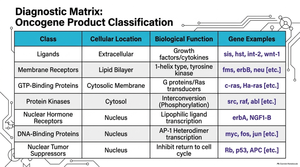
To systematically analyze cancer pathways, biochemists use a comprehensive diagnostic matrix that categorizes signaling components by cellular location and biological function. Classifying these disease-causing factors allows researchers to trace how growth signals travel from the extracellular space into the nucleus. This structured framework groups oncogenes into distinct functional categories, clarifying how specific genetic alterations disrupt cellular balance.
The matrix organizes signaling components across five primary cellular compartments. Extracellular ligands like sis and transmembrane receptors like erbB operate at the outer boundary. Cytosolic membrane switches like ras and cytosolic kinases like src transduce signals through the cytoplasm. Finally, nuclear hormone receptors like ERα, DNA-binding proteins like MYC, and tumor suppressors like p53 govern transcriptional output directly inside the nucleus.
This diagnostic matrix underscores the diverse molecular mechanisms underlying cancer biology. Rather than viewing tumor development as a single, uniform defect, scientists can pinpoint the exact level of regulation that fails. By systematically categorizing these altered factors, medical researchers can design targeted therapies tailored to specific signaling failures. Mapping these functional pathways clarifies how different oncogene classes cooperate to drive disease progression, guiding precision medicine approaches. This matrix serves as an essential roadmap for targeted therapies directed against specific oncogenes across cellular compartments.
Slide 15: The Timeline of Tumorigenesis Driven by Oncogenes
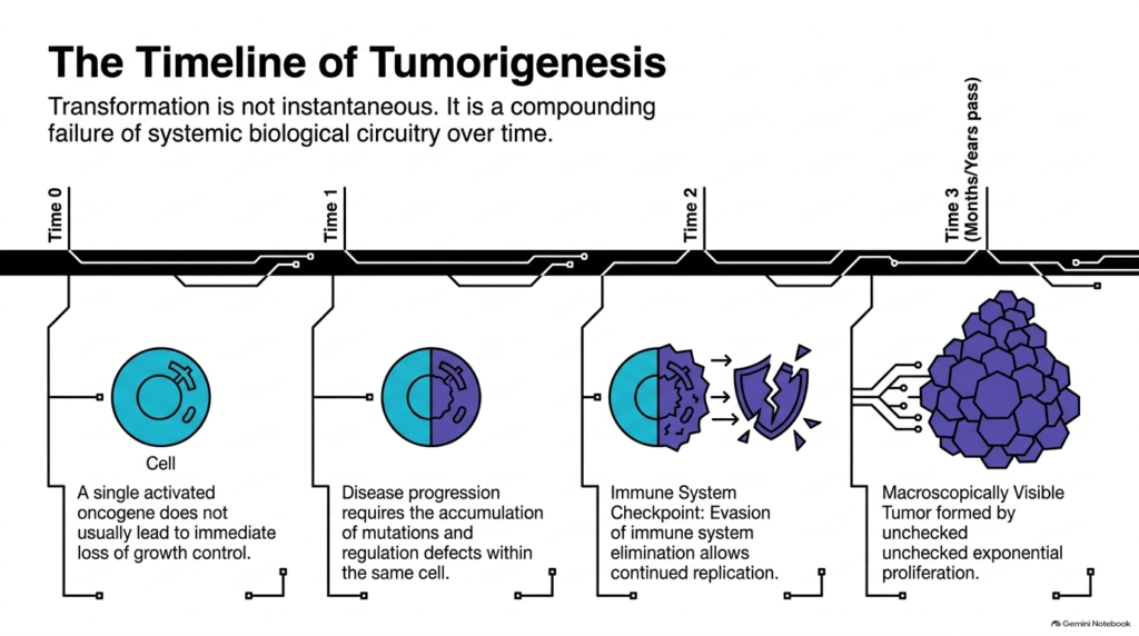
Cellular transformation is never instantaneous; it reflects a compounding failure of complex biological circuitry that unfolds over months or years. Understanding the timeline of tumorigenesis reveals how cells gradually overcome multiple layers of tissue regulation. Throughout this multi-step process, oncogene-driven pathway changes accumulate sequentially, progressively dismantling the cell’s internal safeguards.
At Time 0, a cell acquires an initial mutation, but a single hyperactive gene rarely immediately loses growth control. By Time 1, disease progression requires additional mutations and regulatory defects to accumulate within that same cell lineage. As additional mutations strike, activated oncogenes continuously push the cell toward faster replication, increasing the probability of further genetic errors during DNA division.
By Time 2, the growing clone faces the immune-system checkpoint, where rogue cells must evade immune surveillance and destruction. Successful evasion allows continued replication until, at Time 3, unchecked exponential growth produces a macroscopically visible tumor. This temporal progression highlights that tumorigenesis requires time and multiple genetic hits. By tracking how these oncogenes alter cellular behavior over time, scientists can identify early intervention windows to halt disease progression before tumors mature into invasive cancers. Understanding these temporal dynamics helps clinicians disrupt early oncogenic driver events.
Please read our Content Disclaimer Statement.
Check out our social media channels: