126. Renal Electrolyte Resorption: Mechanisms and Hormonal Regulation
Every single day, human kidneys filter hundreds of liters of blood plasma, producing primary urine packed with vital nutrients, water, and essential ions. If the body excreted this fluid, survival would be impossible within hours. Fortunately, complex cellular machinery constantly recovers these crucial components and returns them to circulation. This blog post breaks down an expert slide deck that examines how tubule cells use energy-dependent transport, hormonal regulation, and metabolic synthesis to preserve internal equilibrium and sustain human life.
Slide 1: Introduction to Renal Electrolyte Resorption and Transport
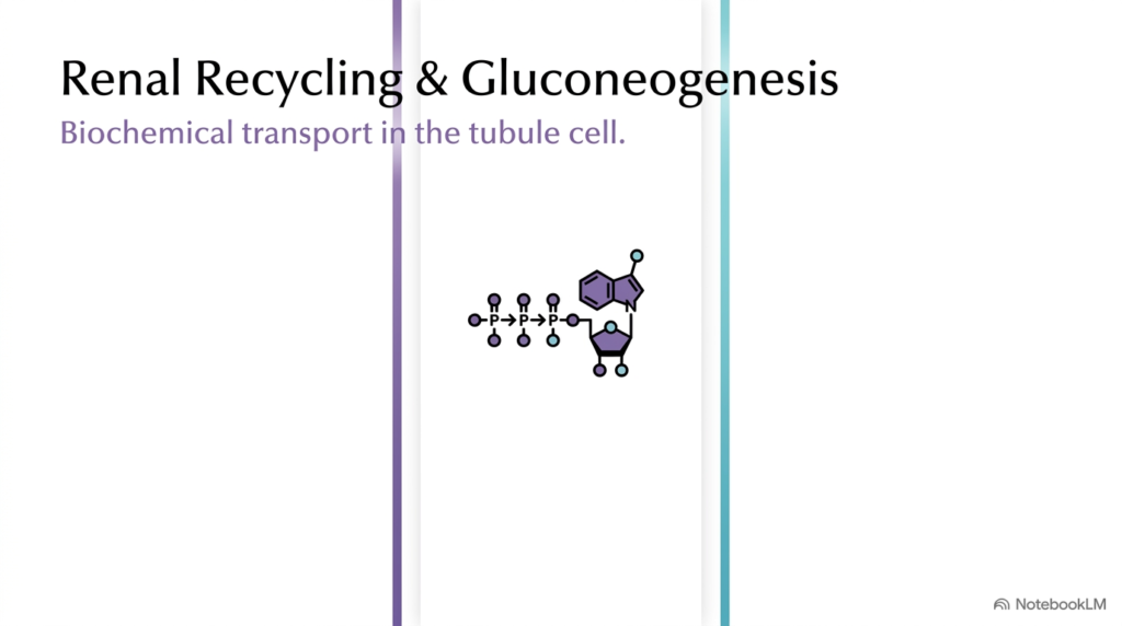
The human kidneys perform a vital role in physiology by constantly filtering blood plasma to create primary urine. Within the microscopic nephron lumen, specialized epithelial tubule cells face the daunting task of sorting through vast volumes of fluid to recover essential solutes before waste leaves the body. This continuous cellular process of Renal Electrolyte Resorption ensures that critical ions, organic nutrients, and water return safely to blood plasma rather than draining away. Hydrolysis of adenosine triphosphate provides the chemical energy that powers active transport pumps across tubule cell membranes.
Beyond reclaiming essential solutes from the primary filtrate, kidney tubule cells show extraordinary metabolic versatility by performing gluconeogenesis alongside active transport. As primary urine flows along the nephron, tubule cells systematically harvest targeted molecules while synthesizing fresh glucose to meet high local cellular energy demands. Understanding Renal Electrolyte Resorption reveals how membrane transport proteins, electrochemical gradients, and metabolic pathways operate in perfect harmony.
Without efficient transport systems, losing essential solutes would rapidly compromise cardiovascular stability and cellular function throughout the body. Effective Renal Electrolyte Resorption provides the physiological foundation for maintaining whole-body fluid balance, blood pressure regulation, and systemic homeostasis. This opening slide introduces the core bioenergetic architecture that enables renal cells to carry out these essential transport and biosynthetic functions.
Slide 2: Three-Zone Architecture of Renal Electrolyte Resorption
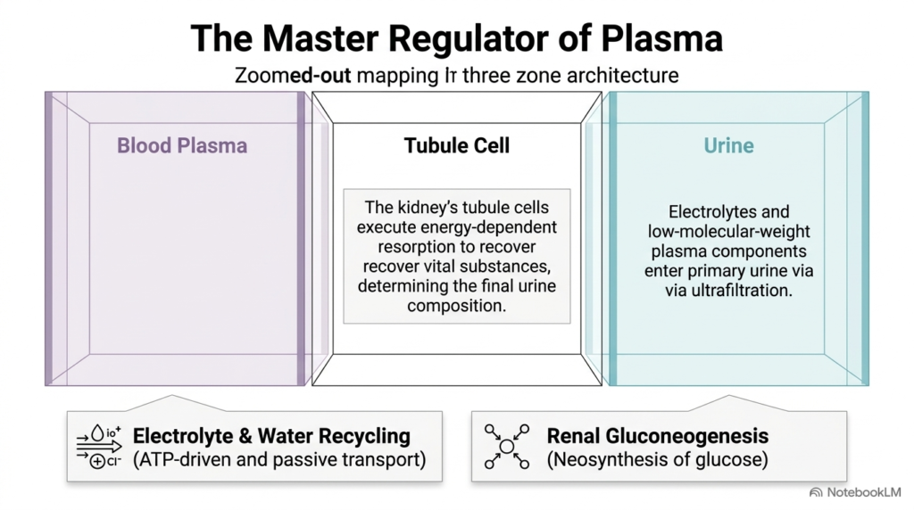
Understanding renal transport mechanisms requires examining the kidney’s structural architecture, organized into three functional zones: blood plasma, tubule cells, and urine. The process begins when blood plasma undergoes ultrafiltration across the glomerular membrane, producing primary urine filled with low-molecular-weight solutes, electrolytes, and water. Tubule cells sit strategically between the urine lumen and surrounding peritubular capillaries, executing energy-dependent transport to reclaim vital compounds. This spatial arrangement establishes the physical framework required for effective Renal Electrolyte Resorption across cellular boundaries.
Within this three-zone architectural model, tubule cells utilize both active and passive transport mechanisms to dictate final urine composition. Energy-driven transcellular pumps actively pull needed ions out of the luminal fluid, while metabolic pathways like renal gluconeogenesis synthesize new glucose molecules internally. By continuously moving targeted solutes from the urine compartment back into peritubular capillaries, Renal Electrolyte Resorption preserves plasma volume and solute concentrations. This elegant organization demonstrates how localized epithelial transport directly governs whole-body internal stability.
Furthermore, the clear separation between urine and blood plasma allows tubule cells to establish steep concentration gradients. As primary urine flows past the apical membrane, active transport mechanisms reabsorb valuable nutrients before they reach the collecting system. Through continuous Renal Electrolyte Resorption, the three-zone architecture prevents catastrophic fluid loss while enabling precise metabolic regulation.
Slide 3: Primary Active Transport in Renal Electrolyte Resorption
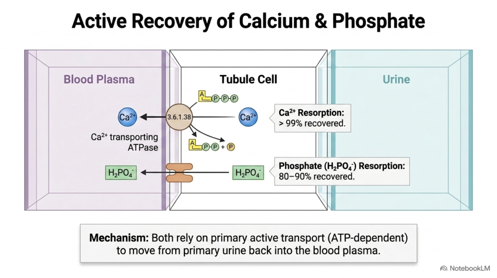
The reclamation of essential divalent ions relies heavily on primary active transport systems embedded within tubule cell membranes. Calcium recovery is remarkably efficient, with cells reclaiming over ninety-nine percent of filtered calcium from primary urine back into blood plasma. Moving calcium against a steep electrochemical gradient requires direct energy input from specialized calcium-transporting ATPases that hydrolyze adenosine triphosphate. Through this dedicated enzymatic mechanism, Renal Electrolyte Resorption maintains optimal systemic calcium levels critical for muscle contraction, nerve conduction, and structural bone integrity.
Phosphate recovery occurs through parallel ATP-dependent active transport pathways, reclaiming eighty to ninety percent of filtered inorganic phosphate as hydrogen phosphate ions. Specific transcellular carrier proteins transport phosphate across the epithelial barrier directly into plasma capillaries. Without continuous energy expenditure, these vital minerals would escape into final urine, producing severe systemic deficiencies. Effective Renal Electrolyte Resorption of calcium and phosphate highlights the absolute necessity of primary active transport engines in sustaining essential mineral balances.
By utilizing primary active transport, tubule cells maintain distinct intracellular environments while moving ions against formidable concentration gradients. Calcium-transporting ATPases keep intracellular calcium extremely low while moving large amounts into circulation. This regulated Renal Electrolyte Resorption of calcium and phosphate demonstrates how energy expenditure directly protects mineral homeostasis.
Slide 4: Hormonal Regulation of Mineral and Renal Electrolyte Resorption
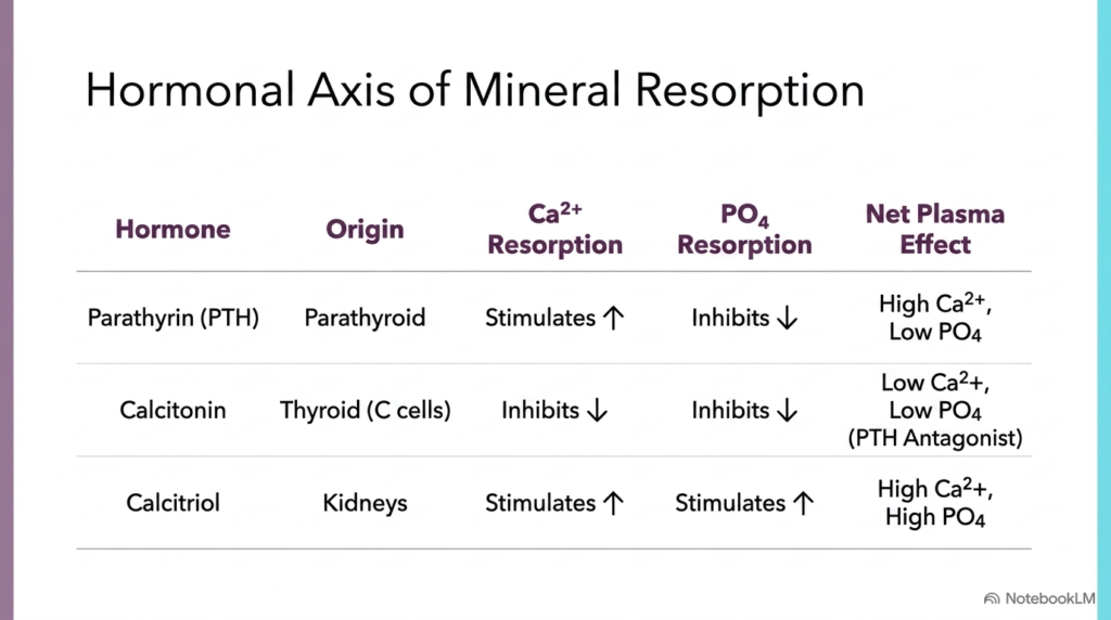
Endocrine signaling controls the exact rate of mineral recovery within kidney tubules to match the body’s shifting physiological demands. Parathyrin, secreted by the parathyroid glands, strongly stimulates calcium reabsorption while inhibiting phosphate reabsorption, resulting in elevated plasma calcium and decreased plasma phosphate concentrations. Conversely, calcitonin released from thyroid C cells acts as a functional antagonist to parathyrin by inhibiting the recovery of both ions. These opposing hormonal signals dynamically adjust Renal Electrolyte Resorption to keep circulating mineral levels within narrow physiological boundaries.
Calcitriol, the active form of vitamin D synthesized within renal tissue, provides powerful complementary regulation by stimulating the reclamation of both calcium and phosphate ions into blood plasma. This dual stimulation boosts systemic mineral availability for bone mineralization and cellular signaling. When endocrine glands detect subtle shifts in blood chemistry, they alter hormone output to fine-tune transport protein activity. Through these coordinated hormonal pathways, Renal Electrolyte Resorption adapts dynamically to defend plasma homeostasis against dietary variations and metabolic challenges.
The interplay between parathyroid hormone, calcitonin, and calcitriol illustrates how systemic feedback loops govern nephron transport proteins. By altering transporter expression and activity in response to hormonal cues, tubule cells fine-tune ion recovery. This multi-hormonal axis governing Renal Electrolyte Resorption demonstrates how endocrine integration prevents mineral toxicity or depletion.
Slide 5: Bulk Sodium Recovery in Renal Electrolyte Resorption
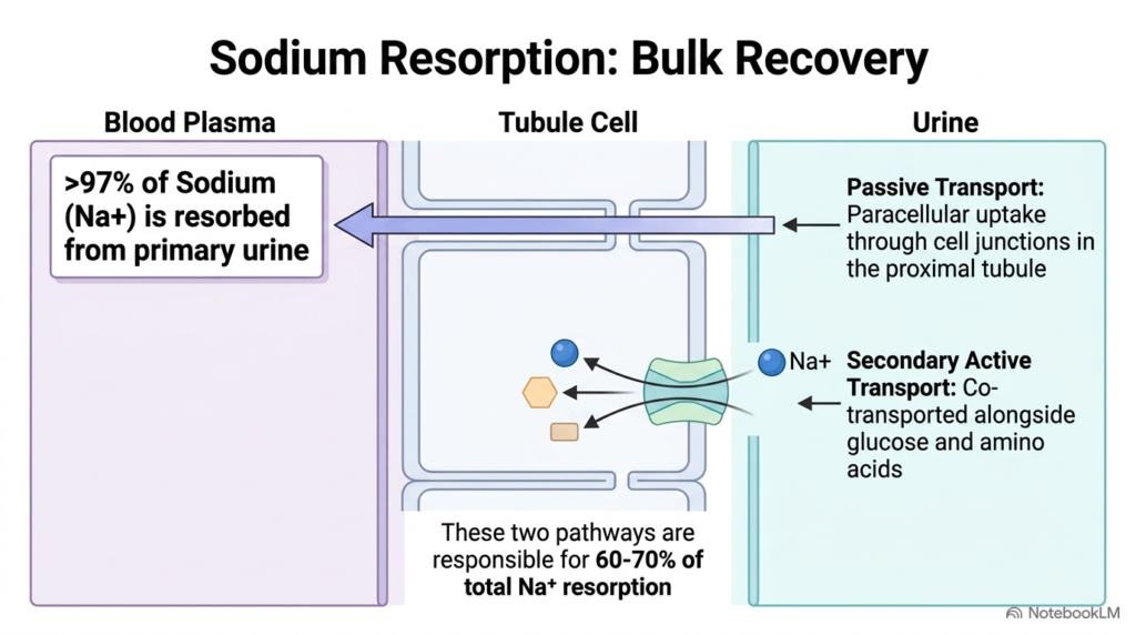
Sodium represents the predominant extracellular cation, making its extensive recovery a top physiological priority for kidney tubule cells. Over 97% of all filtered sodium is reclaimed from primary urine and returned to blood plasma. The proximal tubule handles bulk recovery, reclaiming sixty to seventy percent of total sodium through two distinct transport routes. Passive paracellular transport lets sodium ions diffuse through tight cell junctions, while secondary active transport co-transports sodium with essential nutrients like glucose and amino acids across apical membranes.
Coupling sodium movement to organic nutrient uptake provides a remarkably efficient way to harvest multiple vital substances simultaneously. As sodium moves down its electrochemical gradient into the cell, it drags glucose and amino acids along through specialized symporters. This combined transport strategy ensures rapid bulk solute recovery in early nephron segments. Consequently, Renal Electrolyte Resorption of sodium establishes the primary osmotic driving force that pulls water and secondary solutes back into blood circulation.
Without this high-capacity proximal transport system, the massive volume of filtered sodium would overwhelm distal nephron segments. Proximal tubule cells expend substantial energy to sustain high rates of sodium transport. Through continuous Renal Electrolyte Resorption, the kidney reclaims bulk electrolytes, protecting fluid volume and osmotic balance. Ultimately, effective Renal Electrolyte Resorption in the proximal tubule sets the stage for downstream electrolyte fine-tuning.
Slide 6: ATP-Driven Engines Powering Renal Electrolyte Resorption
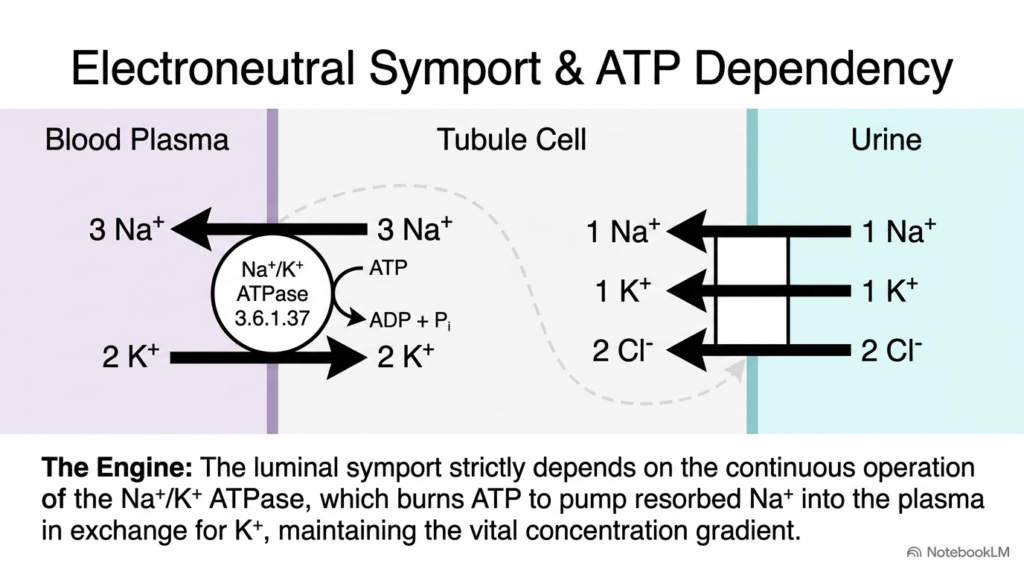
At the cellular level, active solute transport relies on specialized membrane proteins working in precise enzymatic coordination across epithelial borders. On the luminal membrane facing primary urine, an electroneutral symporter imports one sodium ion, one potassium ion, and two chloride ions into the tubule cell simultaneously. This inward movement relies entirely on low intracellular sodium levels established by basolateral pumps. Without continuous primary active transport on the opposite side of the cell, Renal Electrolyte Resorption across the luminal membrane would quickly grind to a halt.
The true engine driving this transport cycle is the basolateral sodium-potassium ATPase, an enzyme that consumes adenosine triphosphate to pump three sodium ions out toward blood plasma while bringing two potassium ions into the cell. By continuously clearing internal sodium, this primary pump preserves the steep electrochemical gradient necessary for luminal symport activity. The seamless integration of basolateral ATP hydrolysis with luminal symport demonstrates how Renal Electrolyte Resorption depends on continuous cellular bioenergetics to maintain ion balance.
Furthermore, the stoichiometry of the luminal symporter ensures electroneutral transport, moving positive and negative charges in equal proportions. This allows tubule cells to accumulate intracellular chloride and potassium without disrupting membrane potential. Through this coordinated enzymatic setup, Renal Electrolyte Resorption efficiently recovers multiple essential electrolytes in a single coupled pathway.
Slide 7: Endocrine Fine-Tuning of Renal Electrolyte Resorption
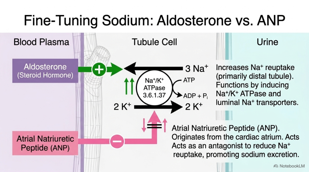
While early nephron segments handle bulk solute recovery, distal tubule segments fine-tune final sodium levels under strict endocrine control. Aldosterone, a steroid hormone released by the adrenal cortex, acts directly on distal tubule cells to enhance sodium reuptake. It does so by inducing synthesis and membrane insertion of basolateral sodium-potassium ATPases and luminal sodium channels. Through this targeted upregulation, Renal Electrolyte Resorption increases significantly, expanding blood volume and raising systemic blood pressure.
In direct opposition to aldosterone, atrial natriuretic peptide released from the cardiac atrium acts as a powerful physiological antagonist to promote sodium excretion. When elevated blood volume stretches heart tissue, atrial natriuretic peptide inhibits sodium transport proteins and suppresses aldosterone action, reducing sodium reuptake. This elegant push-pull endocrine mechanism allows the body to make minute adjustments to sodium recovery. Through these balanced hormonal signals, Renal Electrolyte Resorption precisely regulates blood pressure and extracellular fluid balance.
This hormonal balance ensures that sodium reuptake dynamically responds to cardiovascular pressures and fluid volume states. When dehydrated, aldosterone levels surge to maximize sodium retention, whereas fluid overload triggers atrial natriuretic peptide release. By fine-tuning nephron transport, Renal Electrolyte Resorption maintains optimal blood pressure and extracellular solute composition under changing environmental conditions.
Slide 8: Water Dynamics and Osmotic Renal Electrolyte Resorption
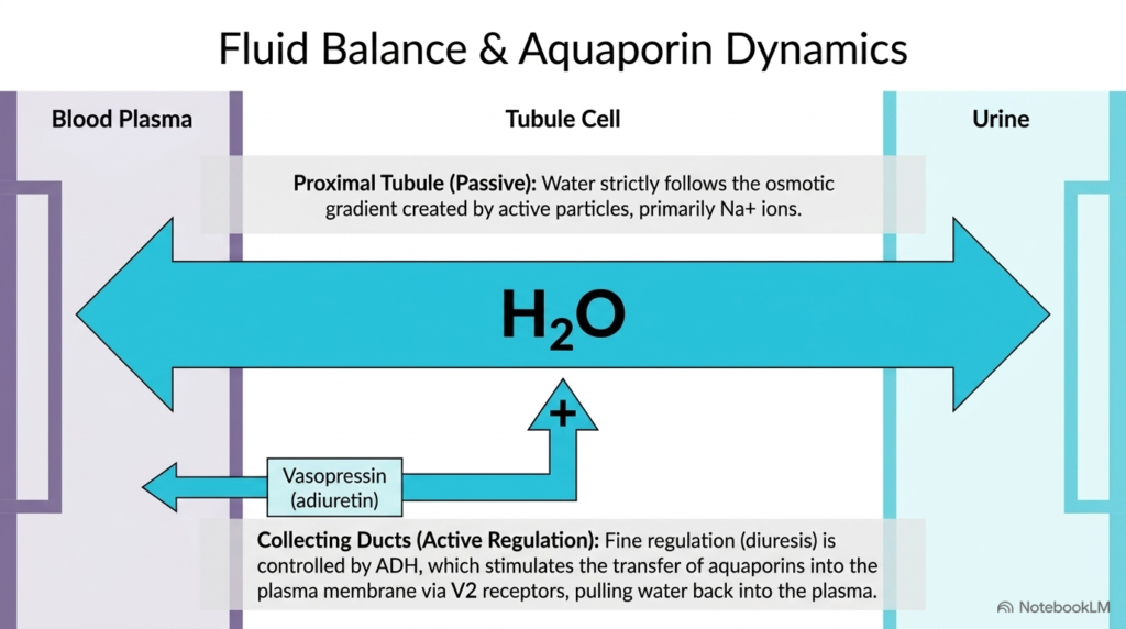
Water recovery in the kidney is intimately tied to the movement of dissolved ions across epithelial barriers. In the proximal tubule, water reabsorption occurs passively as water molecules spontaneously follow the osmotic gradient generated by active sodium movement. Where sodium goes, water naturally follows through highly permeable cell membranes. This obligatory fluid movement ensures that large volumes of water return to blood plasma alongside solutes, demonstrating how Renal Electrolyte Resorption directly creates the osmotic force required for fluid balance.
In contrast to obligatory proximal uptake, water recovery in the collecting ducts is strictly hormonally regulated to determine final urine concentration. Vasopressin, also known as antidiuretic hormone, binds to V2 receptors on tubule cells, triggering intracellular signaling cascades that insert aquaporin water channels into the plasma membrane. These specialized channels allow water to leave the urine and re-enter circulation rapidly. Through vasopressin-mediated aquaporin dynamics, Renal Electrolyte Resorption couples solute transport to precise regulation of whole-body hydration.
Without aquaporin insertion, collecting duct walls remain impermeable to water, leading to dilute urine excretion. Vasopressin acts as a molecular switch, allowing tubule cells to adjust water permeability on demand. Through this sophisticated mechanism, Renal Electrolyte Resorption and regulated aquaporin channels work together to defend plasma osmolarity against severe dehydration.
Slide 9: Clinical Pathology Impairing Renal Electrolyte Resorption
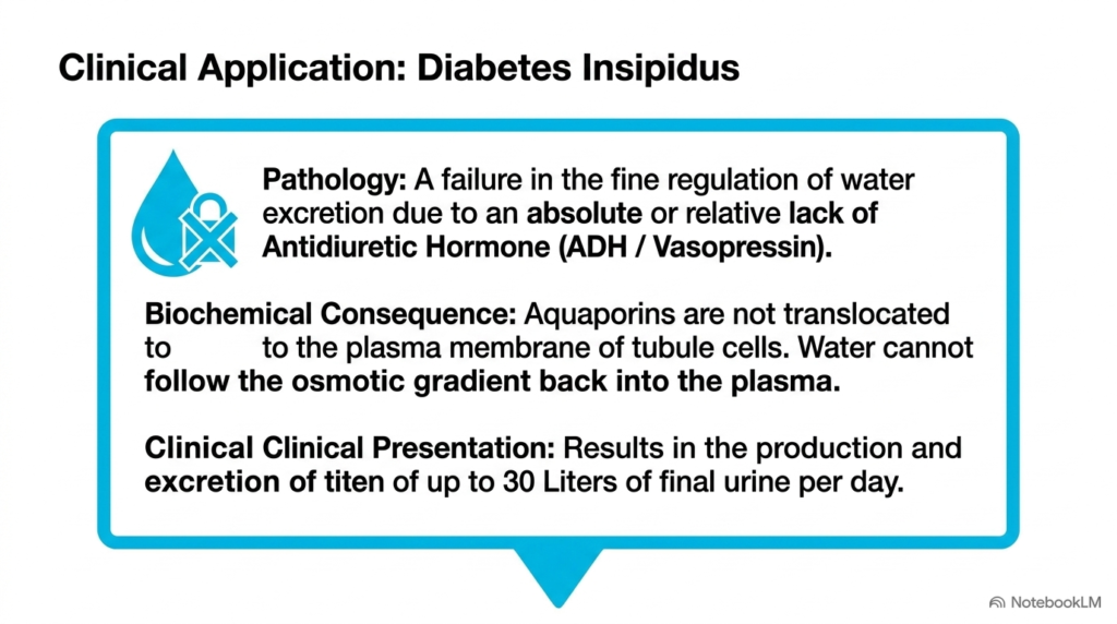
Disruptions in renal transport mechanisms can produce profound clinical disorders that severely threaten systemic health. Diabetes insipidus represents a classic pathology marked by a breakdown in the fine regulation of water excretion. This condition stems from an absolute deficiency of vasopressin production or a relative resistance to its action at the kidney tubule level. When vasopressin signaling fails, tubule cells cannot translocate aquaporin channels into their plasma membranes, preventing water from following osmotic gradients back into blood plasma.
Without functional aquaporin channels in collecting duct membranes, massive volumes of diluted fluid pass unchecked into final urine. Patients suffering from severe diabetes insipidus can excrete up to thirty liters of urine daily, leading to rapid dehydration and dangerous electrolyte imbalances. This clinical presentation highlights how impaired fluid recovery secondary to defective Renal Electrolyte Resorption destabilizes whole-body fluid homeostasis. Understanding these cellular defects underscores the clinical importance of intact hormonal signaling in tubular transport pathways.
The inability to reabsorb water leads to severe hypernatremia and intense thirst as the body desperately attempts to compensate for urinary losses. Clinical treatment requires administering exogenous vasopressin analogs or addressing receptor resistance. This severe disorder illustrates how critical intact Renal Electrolyte Resorption and aquaporin translocation are for maintaining life-sustaining fluid balance, showing that proper Renal Electrolyte Reabsorption is essential for patient survival.
Slide 10: Metabolic Coupling of Gluconeogenesis and Renal Electrolyte Resorption
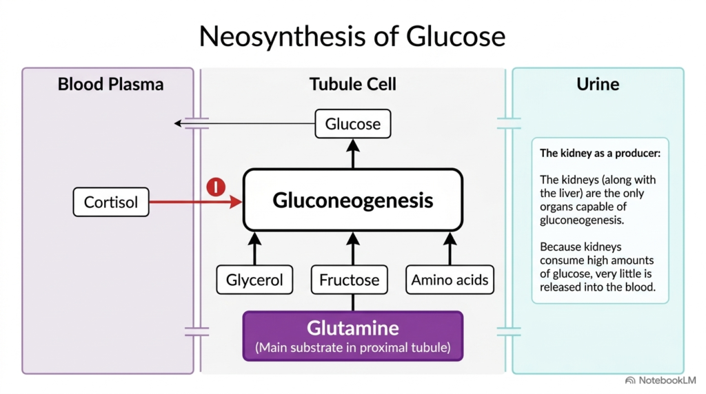
In addition to solute transport, kidney tubule cells have the unique metabolic capability to synthesize glucose from non-carbohydrate precursors. Along with the liver, the kidneys are the only organs that can perform gluconeogenesis to generate new glucose molecules. Proximal tubule cells utilize glycerol, fructose, amino acids, and primarily glutamine as metabolic substrates for this biosynthetic pathway. Glucocorticoids like cortisol regulate this process, aligning glucose production with systemic metabolic stress and energy demands in renal tissue.
Renal gluconeogenesis is functionally integrated with tubular transport activities, providing local metabolic energy while supporting plasma glucose levels. Because tubule cells consume substantial glucose to power active ion pumps, they use much of the newly synthesized sugar locally rather than releasing it into circulation. This tight metabolic integration demonstrates how energy production directly fuels active transport processes. Ultimately, Renal Electrolyte Resorption and glucose neosynthesis operate side by side to sustain cellular viability and metabolic equilibrium.
Furthermore, glutamine breakdown during gluconeogenesis yields carbon skeletons for energy while releasing nitrogenous byproducts. Cortisol stimulation enhances both enzyme activity and substrate uptake in proximal tubule cells during fasting or stress. Through this dual function, Renal Electrolyte Resorption and renal gluconeogenesis collaborate to supply metabolic fuel, showing that continuous Renal Electrolyte Reabsorption relies on robust intracellular energy production.
Slide 11: Acid-Base Buffering Integrated with Renal Electrolyte Resorption
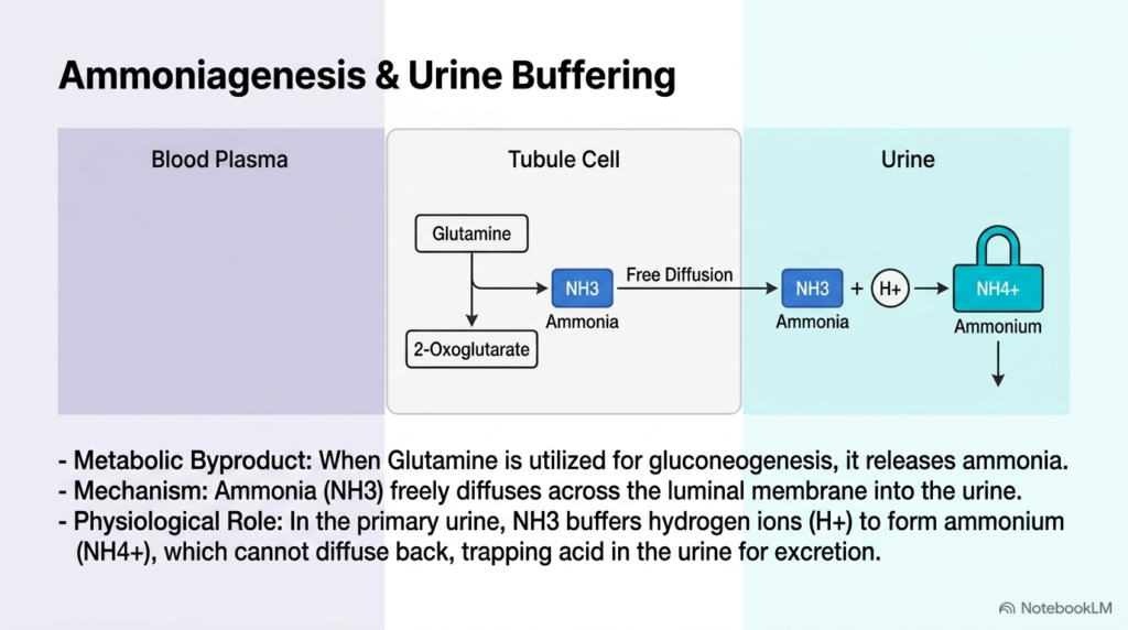
Maintaining systemic blood pH requires kidney tubule cells to coordinate metabolic pathways with urinary acid excretion. When tubule cells utilize glutamine for gluconeogenesis, the breakdown of this amino acid generates ammonia alongside alpha-ketoglutarate as a metabolic byproduct. Uncharged ammonia molecules diffuse freely across luminal cell membranes into primary urine. This enzymatic production of ammonia provides a continuous supply of urinary buffer, enabling the kidney to eliminate excess metabolic hydrogen ions without drastically lowering urine pH.
Once inside primary urine, free ammonia reacts with hydrogen ions to form ammonium ions, which carry a positive charge. Because charged ammonium cannot diffuse back across cell membranes into tubule cells, acid becomes permanently trapped in urine for excretion. This biochemical trapping mechanism allows the body to clear metabolic acid efficiently while recovering valuable bicarbonate. Through this elegant integration of ammoniagenesis and solute transport, Renal Electrolyte Resorption plays a crucial role in defending systemic acid-base balance.
This urinary buffering system prevents damaging acidic conditions in tubular fluid while allowing continuous proton excretion. By coupling glutamine breakdown to acid elimination, tubule cells protect systemic pH during metabolic acidosis. Through these integrated biochemical pathways, Renal Electrolyte Resorption supports both acid elimination and essential electrolyte preservation, demonstrating how Renal Electrolyte Resorption links metabolic buffering directly to ion conservation.
Please read our Content Disclaimer Statement.
Check out our social media channels: