148. Steroid Hormone Metabolism: Biosynthesis, Pathways, and Inactivation
Have you ever wondered how a single chemical precursor transforms into signals that regulate your stress response, muscle growth, and fluid balance? Steroid Hormone Metabolism is the master cellular pathway that creates these diverse signaling molecules. This slide deck delivers a step-by-step breakdown of how cells synthesize, structure, and clear these vital lipid signals. Designed for college and medical school students, this comprehensive visual guide distills complex metabolic cascades into clear biochemical principles.
Slide 01: Steroid Hormone Metabolism – Core Pathways and Chemical Foundations
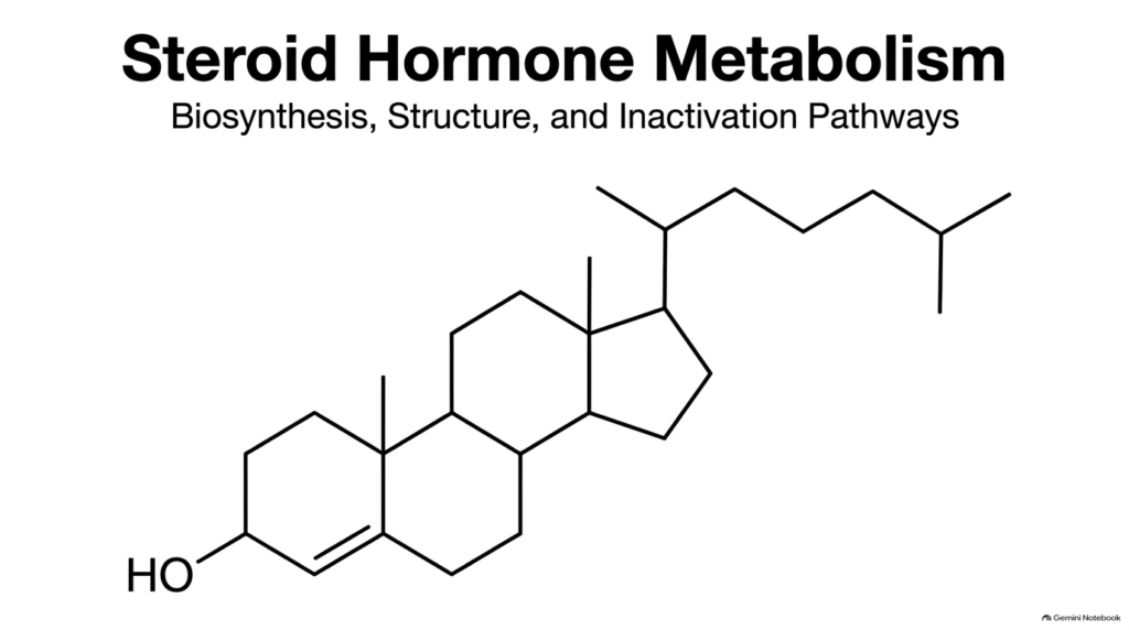
Studying steroid hormone metabolism begins with understanding how complex lipophilic signals govern systemic physiology across human tissues. This introductory slide establishes the core framework connecting dietary lipid precursors to highly active signaling compounds. Without tightly regulated steroid hormone metabolism, target tissues would completely lack the essential chemical messengers needed to coordinate cellular homeostasis, reproductive cycles, and acute stress responses throughout the organism.
At its structural foundation, steroid hormone metabolism relies on a shared gonane nucleus composed of four fused carbon rings labeled A, B, C, and D. This specific arrangement of seventeen carbon atoms forms the universal chemical scaffold for all major endocrine steroid classes. Glandular cells systematically modify this hydrophobic core through targeted enzymatic reactions to generate distinct hormonal capabilities. By visualizing this initial parent structure, students can easily appreciate how subtle shifts in functional groups dramatically alter biological affinity and receptor selectivity.
Throughout the human endocrine network, specialized cells execute these precise structural changes to maintain healthy physiological output. Mastering steroid hormone metabolism requires recognizing how precursor molecules transition between organelle compartments to reach their final active state. This opening visual sets an essential foundation for exploring the multi-step cascades that transform basic cholesterol into potent regulators and prepare them for eventual hepatic elimination.
Slide 02: Steroid Hormone Metabolism – Cholesterol as the Universal Precursor
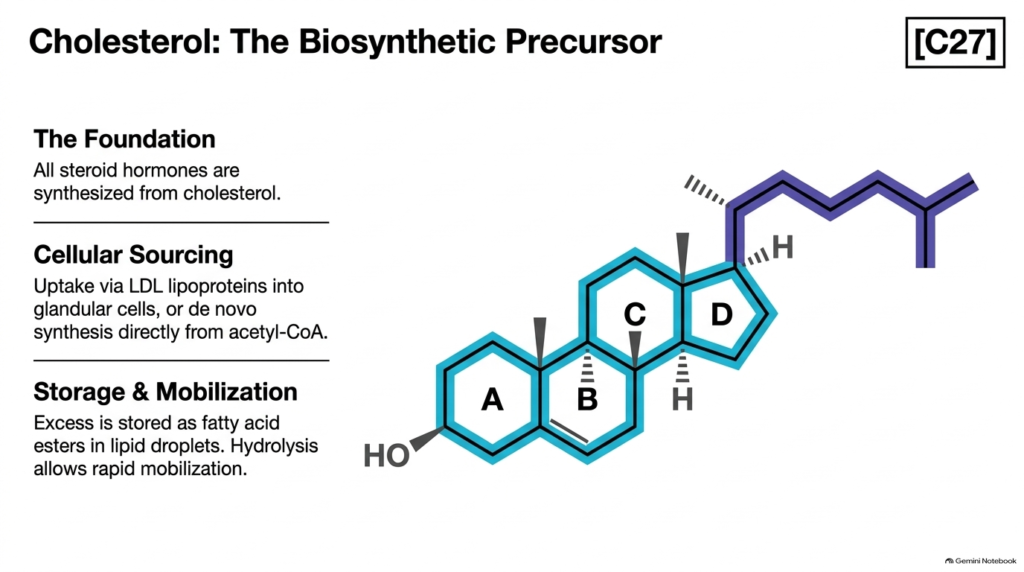
Every biochemical pathway in steroid hormone metabolism relies entirely on cholesterol as its foundational parent molecule. Containing twenty-seven carbon atoms arranged in four fused hydrocarbon rings with an eight-carbon side chain, cholesterol provides the essential hydrophobic framework for all steroid biosynthesis. Glandular cells efficiently acquire this precursor through two distinct cellular routes: absorbing circulating low-density lipoproteins via receptor-mediated endocytosis or synthesizing cholesterol directly from acetyl-CoA units.
To ensure a continuous substrate supply for steroid hormone metabolism, endocrine cells maintain specialized lipid storage pools. Cells esterify excess cholesterol with fatty acids and store these hydrophobic molecules safely inside cytosolic lipid droplets. When physiological signals trigger acute hormone demand, intracellular esterases rapidly hydrolyze these ester bonds, liberating free cholesterol for immediate transport into mitochondria. This mobilization step acts as a critical regulatory checkpoint that directly dictates downstream metabolic activity.
Understanding this initial substrate stage is fundamental to mastering overall steroid hormone metabolism. The instant availability of unesterified cholesterol determines how effectively endocrine tissues respond to systemic stimuli. By examining how glandular cells harvest, store, and mobilize this twenty-seven-carbon sterol, students gain vital insight into the primary cellular investment required before specialized glucocorticoids, mineralocorticoids, or sex steroids can be constructed.
Slide 03: Steroid Hormone Metabolism – The Specialized Enzymatic Toolkit
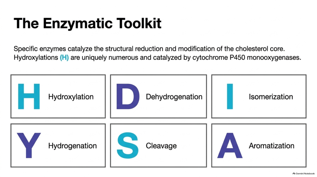
The structural progression of steroid hormone metabolism depends on a versatile toolkit of six primary enzymatic reactions. Endocrine cells transform the rigid cholesterol skeleton through precise chemical modifications that alter functional groups to dictate biological receptor specificity. Hydroxylations are the most common reaction type in steroid hormone metabolism, introducing polar hydroxyl groups at specific carbon positions. These crucial oxygenations are catalyzed by specialized monooxygenases belonging to the cytochrome P450 superfamily.
Beyond hydroxylation, active steroid hormone metabolism relies on dehydrogenation and hydrogenation reactions to modify molecular oxidation states. Pyridine nucleotide-dependent dehydrogenases oxidize hydroxyl groups to keto functions or shift double-bond positions, whereas NADPH-dependent hydrogenases reduce double bonds through targeted hydrogen addition. Isomerases further assist by shifting double bonds between adjacent carbon atoms, successfully reorganizing ring chemistry to stabilize reactive intermediates during complex synthetic sequences.
The catalytic toolset governing steroid hormone metabolism is completed by carbon cleavage enzymes and aromatase. Lyase-mediated cleavage reactions shorten or entirely eliminate bulky carbon side chains to systematically alter overall carbon counts. Meanwhile, aromatase uniquely transforms saturated carbon rings into stable aromatic structures. Mastering these six fundamental catalytic mechanisms lets students predict structural outcomes and functional changes across every steroid hormone lineage.
Slide 04: Steroid Hormone Metabolism – The Carbon Reduction Cascade
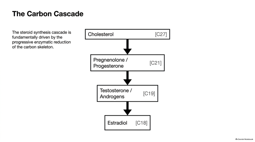
The overarching architecture of steroid hormone metabolism follows a logical, highly organized carbon reduction cascade. Rather than building larger molecules, steroidogenesis systematically trims the parent cholesterol skeleton into smaller, highly specialized endocrine signals. This progressive reduction in overall carbon count is a primary organizing principle throughout steroid hormone metabolism, allowing students to classify steroid classes cleanly based on their precise carbon frame.
The pathway begins with twenty-seven-carbon cholesterol and steps down through distinct structural tiers during steroid hormone metabolism. Initial side-chain cleavage reduces the skeleton to twenty-one carbons, forming essential gestagens, glucocorticoids, and mineralocorticoids such as progesterone, cortisol, and aldosterone. Subsequent enzymatic removal of two additional carbons from the core generates nineteen-carbon androgens such as testosterone and androstenedione. Finally, losing the C-19 angular methyl group produces eighteen-carbon estrogens.
Visualizing this sequential carbon loss provides a powerful mental map for navigating complex steroid hormone metabolism. Each step down the carbon cascade alters receptor affinity while creating unique structural features for target cell nuclear signaling. By tracking carbon numbers systematically from C27 down through C21, C19, and C18, students can easily trace how specific cleavage events transform a single lipid substrate into distinct physiological regulators that govern diverse human metabolic and developmental functions.
Slide 05: Steroid Hormone Metabolism – Pregnenolone Synthesis and Side-Chain Cleavage
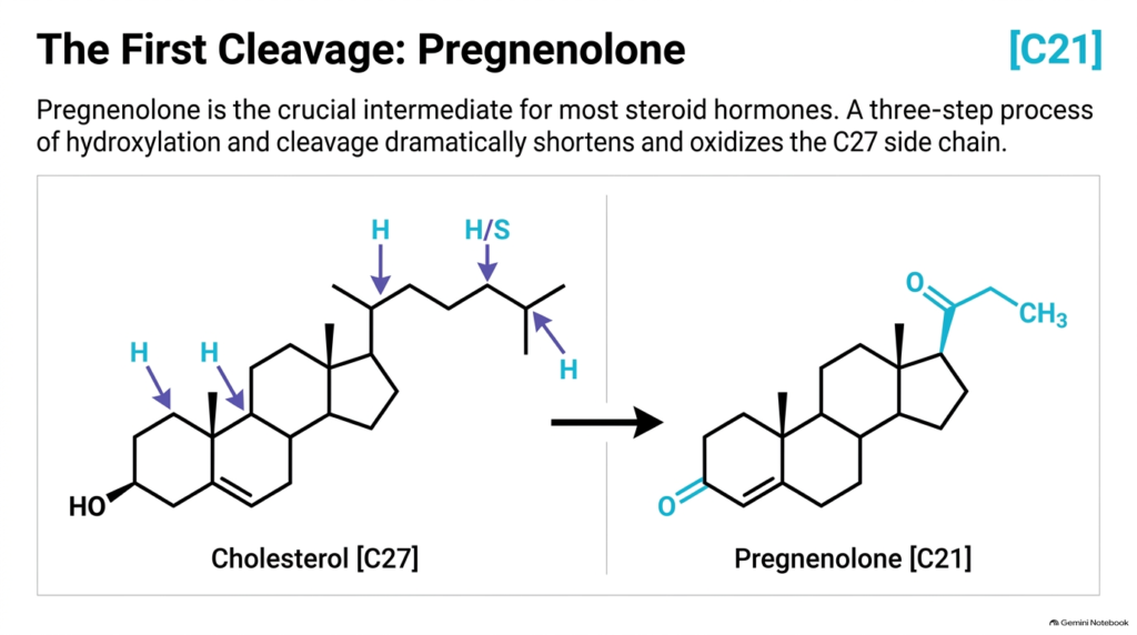
Committed steroid hormone metabolism begins with the conversion of cholesterol to pregnenolone. This pivotal reaction represents the first major carbon-cleavage step in steroidogenesis, transforming the bulky twenty-seven-carbon sterol into a versatile twenty-one-carbon intermediate. Pregnenolone serves as the indispensable intermediate precursor for nearly all functional steroid hormones, making this side-chain cleavage reaction a crucial regulatory milestone in overall steroid hormone metabolism.
Mechanistically, this transformation requires a three-step enzymatic sequence catalyzed by the mitochondrial cytochrome P450 side-chain cleavage enzyme complex. The enzyme executes two consecutive hydroxylations on the hydrocarbon side chain at carbon positions 20 and 22, generating a vicinal diol intermediate. Subsequent oxidative cleavage breaks the carbon-carbon bond between C-20 and C-22, releasing isocaproaldehyde and yielding C21 pregnenolone. This oxidative modification permanently alters the steroid’s hydrophobic character.
Understanding this initial cleavage reaction is essential for mastering steroid hormone metabolism. Because side-chain cleavage occurs exclusively inside the inner mitochondrial membrane, transporting free cholesterol across organelle membranes is the primary rate-limiting step in the pathway. By examining how pregnenolone is formed, students see how glandular cells overcome structural barriers to initiate downstream hormone production, establishing the baseline substrate pool required for all subsequent endocrine synthesis.
Slide 06: Steroid Hormone Metabolism – Progesterone as the Universal Branch Point
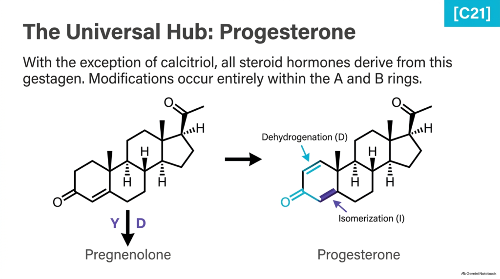
Following initial pregnenolone formation, steroid hormone metabolism advances through the crucial generation of progesterone. Progesterone sits at the center as the universal biosynthetic hub from which all major glucocorticoids, mineralocorticoids, and sex steroids derive. With the sole exception of calcitriol, every classical steroid hormone pathway flows through progesterone, highlighting its indispensability in human steroid hormone metabolism across adrenal and gonadal tissues.
The conversion of pregnenolone into progesterone involves two coordinated catalytic actions occurring within the steroid A and B rings. First, a 3beta-hydroxysteroid dehydrogenase oxidizes the polar hydroxyl group at carbon-3 into a keto group. Next, a steroid isomerase shifts the double bond from C5-C6 to C4-C5, creating a conjugated alpha,beta-unsaturated ketone in ring A. These specific structural modifications significantly stabilize the steroid core, preparing it for downstream oxygenation reactions.
Recognizing progesterone’s pivotal role is essential for comprehending overall steroid hormone metabolism. Because progesterone sits directly at the major biosynthetic crossroads, tissue-specific enzymatic choices at this stage dictate whether precursor flux is directed toward metabolic stress regulation, renal fluid balance, or reproductive maturation. By mastering how progesterone is constructed and modified, students can easily track how different endocrine tissues allocate substrate pathways toward specific physiological and clinical outcomes.
Slide 07: Steroid Hormone Metabolism – Glucocorticoid Biosynthesis and Cortisol
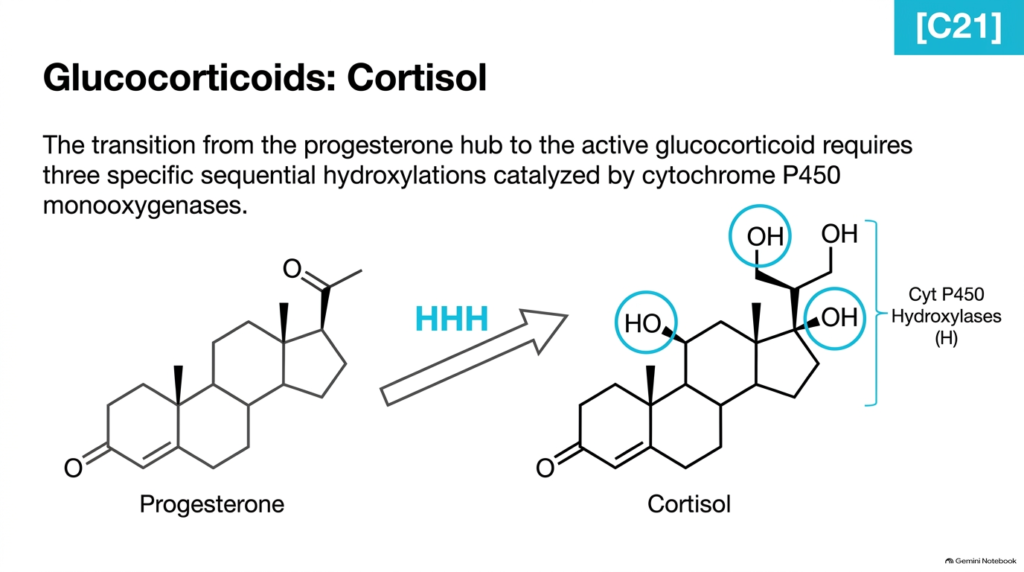
Glucocorticoid synthesis is a major physiological pathway in adrenal steroid hormone metabolism. Cortisol, the principal human glucocorticoid, regulates systemic glucose homeostasis, suppresses immune responses, and supports acute cellular stress adaptation. To transition successfully from the progesterone hub to biologically active cortisol, the adrenal cortex executes three sequential hydroxylation reactions, demonstrating the precise spatial and chemical regioselectivity that characterizes steroid hormone metabolism in adrenal tissues.
Specific cytochrome P450 monooxygenases catalyze these three hydroxylations in distinct adrenal organelle compartments. First, 17alpha-hydroxylase introduces a hydroxyl group at carbon-17 within the endoplasmic reticulum. Next, 21-hydroxylase adds a second hydroxyl group at carbon-21. Finally, the intermediate translocates back into the mitochondria, where 11-beta-hydroxylase adds the third hydroxyl group at carbon-11. This sequential addition of three polar hydroxyl groups transforms hydrophobic progesterone into potent, soluble cortisol.
Understanding cortisol synthesis provides indispensable insight into overall steroid hormone metabolism. The spatial division of enzymes between the endoplasmic reticulum and mitochondrial matrix emphasizes the complex intracellular trafficking required during hormone assembly. By following these three targeted oxygenation steps, students see how subtle chemical modifications dramatically alter receptor specificity, enabling cortisol to selectively bind glucocorticoid receptors and regulate human energy metabolism and inflammatory responses.
Slide 08: Steroid Hormone Metabolism – Mineralocorticoid Synthesis and Aldosterone
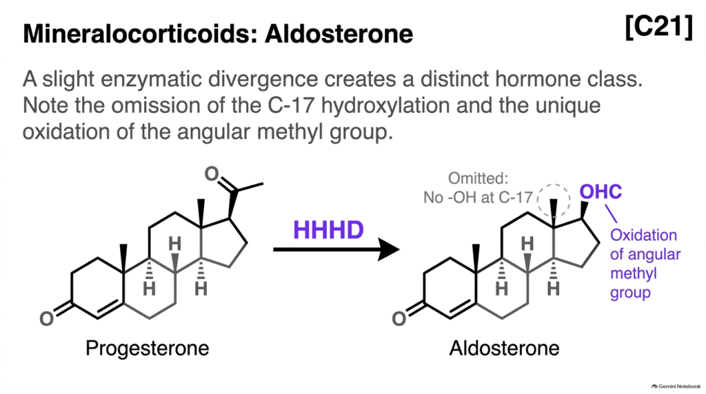
The mineralocorticoid pathway highlights how minor enzymatic shifts in steroid hormone metabolism yield completely different endocrine functions. Aldosterone, produced in the zona glomerulosa of the adrenal cortex, is the chief regulator of renal sodium retention, potassium excretion, and blood pressure control. By altering specific steps in cortisol synthesis, steroid hormone metabolism generates a distinct hormone class tailored to systemic electrolyte balance and plasma volume homeostasis.
Aldosterone biosynthesis diverges from cortisol production at two key enzymatic junctions. First, the 17-alpha-hydroxylation step is completely omitted, keeping carbon-17 unoxygenated. Instead, progesterone undergoes sequential hydroxylations at carbon-21 and carbon-11. The unique hallmark of aldosterone synthesis occurs at carbon-18, where aldosterone synthase oxidizes the angular methyl group into an aldehyde group. This aldehyde group can reversibly react with the nearby C-11 hydroxyl group to form a stable cyclic hemiacetal structure in aqueous environments.
Analyzing aldosterone synthesis reinforces a foundational rule of steroid hormone metabolism: small structural alterations dictate receptor selectivity. Leaving carbon-17 unhydroxylated while oxidizing carbon-18 prevents aldosterone from activating glucocorticoid receptors while maximizing its affinity for mineralocorticoid receptors. Studying this pathway shows students how subtle enzymatic adjustments allow cells to separate renal salt regulation from metabolic energy control across different target tissues.
Slide 09: Steroid Hormone Metabolism – Androgen Formation and Side-Chain Removal
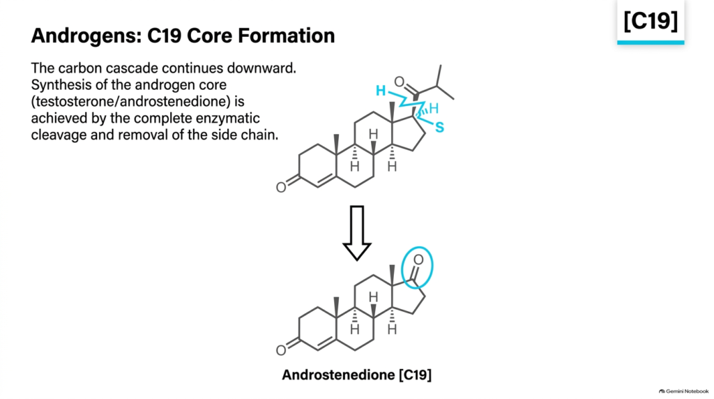
The transition to androgen production marks another major structural milestone in steroid hormone metabolism. Androgens, including testosterone and androstenedione, serve as essential male sex steroids and vital intermediate precursors for estrogen synthesis. To generate these nineteen-carbon molecules, endocrine cells carry out a side-chain cleavage reaction that removes two carbons from the C21 steroid skeleton, showing how steroid hormone metabolism systematically reduces molecular size to shift functional capabilities across tissues.
This carbon removal is catalyzed by the dual-function enzyme 17alpha-hydroxylase/17,20-lyase. First, the 17alpha-hydroxylase activity adds a hydroxyl group at carbon-17 of the C21 substrate. Next, the 17,20-lyase activity cleaves the C17-C20 carbon-carbon bond, completely removing the remaining two-carbon side chain. This cleavage leaves a keto group at carbon-17, forming the C19 androgen core structure typified by androstenedione. Subsequent 17beta-hydroxysteroid dehydrogenase activity reduces this keto group to yield active testosterone.
Understanding androgen synthesis provides a crucial conceptual bridge in steroid hormone metabolism. The complete removal of the side chain fundamentally alters hormone function, shifting biological activity away from corticosteroid signaling toward anabolic growth and reproductive signaling. By analyzing how cells construct the C19 androgen frame, students gain clear insight into the biochemical transitions that separate adrenal steroid production from specialized gonadal sex hormone production during human development.
Slide 10: Steroid Hormone Metabolism – Estrogen Biosynthesis and Aromatization
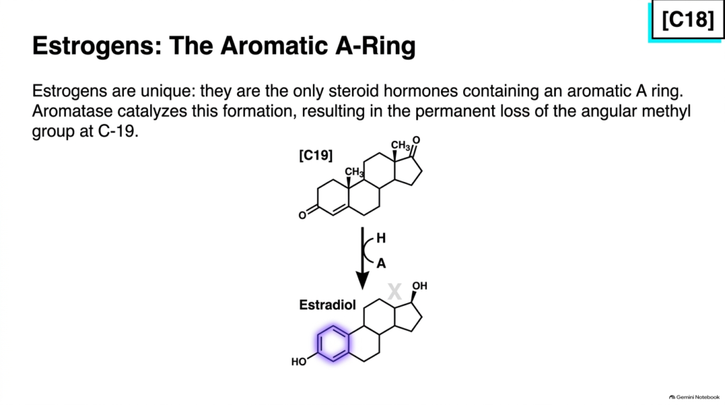
The final step in the synthesis of classic sex steroids represents the most drastic structural change in steroid hormone metabolism. Estrogens, such as estradiol and estrone, are eighteen-carbon steroids characterized by a unique aromatic A ring. Unlike all other steroid hormones, estrogens completely lack the angular C-19 methyl group, illustrating how steroid hormone metabolism eliminates nuclear methyl groups to create distinct female sex hormones with high biological potency across targeted human tissues.
Aromatase, a specialized cytochrome P450 enzyme complex, catalyzes this transformation by converting C19 androgens into C18 estrogens. Aromatase performs three sequential oxidation steps on the C-19 angular methyl group and ring A. The reaction removes carbon-19 as formic acid while simultaneously abstracting hydrogens from ring A to generate a fully aromatic phenolic ring structure. This aromatization step converts testosterone directly into estradiol or androstenedione into estrone.
Mastering estrogen biosynthesis completes the biosynthetic overview of steroid hormone metabolism. Aromatization radically changes the molecule’s physical and chemical properties, making ring A planar and phenolic and enabling selective, high-affinity binding to nuclear estrogen receptors. By recognizing how aromatase converts male sex steroids into female sex steroids, students appreciate the critical enzymatic bridge connecting androgen and estrogen physiology during health and disease.
Slide 11: Steroid Hormone Metabolism – Calcitriol Synthesis and Secosteroid Pathways
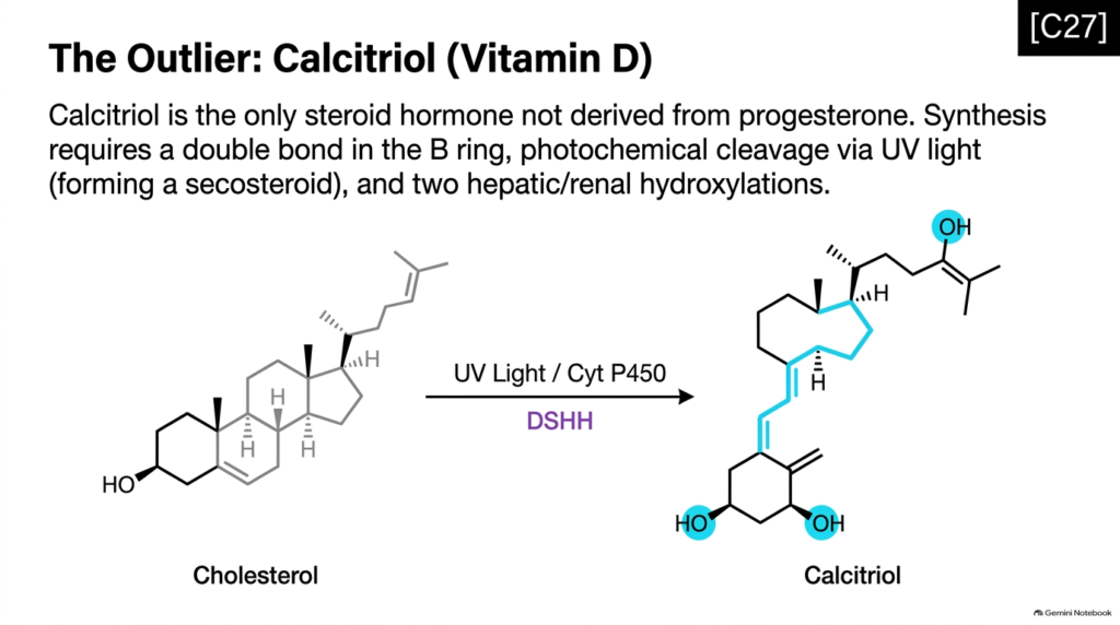
While most classic steroid hormones derive from progesterone, calcitriol stands out as a unique structural outlier in steroid hormone metabolism. Calcitriol, the biologically active form of vitamin D3, retains all twenty-seven carbons of the original cholesterol skeleton and acts as the chief regulator of systemic calcium and phosphate homeostasis. Studying calcitriol reveals an unusual branch of steroid hormone metabolism that integrates environmental photochemical reactions with sequential enzymatic modifications across multiple distinct organs.
Calcitriol synthesis begins in the skin, where 7-dehydrocholesterol undergoes photochemical cleavage under solar ultraviolet B radiation. Sunlight ruptures the B ring between carbon-9 and carbon-10, transforming the intact steroid ring into a broken-ring secosteroid known as cholecalciferol. Cholecalciferol then travels through the bloodstream and undergoes two sequential cytochrome P450-dependent hydroxylations: first at carbon-25 in the liver, then at carbon-1 in the kidneys to produce active calcitriol.
Including calcitriol expands student understanding of steroid hormone metabolism. It demonstrates that potent steroid signaling molecules can arise through ring-opening photochemical steps rather than traditional multi-enzyme side-chain cleavage cascades alone. By mapping calcitriol synthesis across the skin, liver, and kidneys, students learn how organ cooperation and light energy combine to produce vital endocrine regulators essential for human skeletal health and systemic mineral balance.
Slide 12: Steroid Hormone Metabolism – Adrenogenital Syndrome and Enzyme Defects
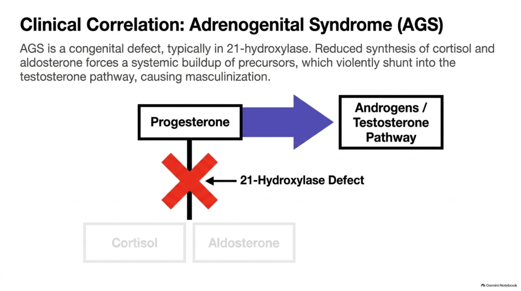
Clinical pathologies provide powerful real-world examples of how disruption in steroid hormone metabolism dramatically alters human physiological development. Adrenogenital syndrome, also known as congenital adrenal hyperplasia, results from inherited enzymatic defects in adrenal steroid synthesis pathways. Examining this clinical condition demonstrates how a single enzyme blockage disrupts the precise balance of steroid hormone metabolism, redirecting accumulated precursor flux into unintended hormonal pathways with profound systemic consequences.
Most adrenogenital syndrome cases stem from a deficiency in 21-hydroxylase, the essential enzyme that synthesizes cortisol and aldosterone from progesterone precursors. When 21-hydroxylase activity is impaired, adrenal cortisol production drops dramatically. Because low cortisol levels eliminate normal negative feedback inhibition on the pituitary gland, adrenocorticotropic hormone secretion spikes significantly. This intense hormonal drive forces massive precursor accumulation, shunting excess progesterone and 17-hydroxyprogesterone into the unimpaired androgen pathway and producing excessive testosterone.
Studying adrenogenital syndrome highlights the deeply interconnected nature of steroid hormone metabolism. The resulting androgen excess causes severe masculinization in female fetuses and precocious puberty in young males. By understanding how upstream enzymatic blockages force metabolic pathway shunting, students see how biochemical pathway regulation directly impacts clinical endocrinology, prenatal diagnostic screening, and early therapeutic hormone interventions before birth.
Slide 13: Steroid Hormone Metabolism – Phase I Hepatic Inactivation
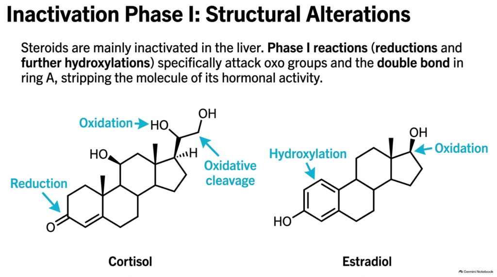
Maintaining proper endocrine balance requires not only efficient hormone synthesis but also rapid clearance mechanisms. Phase I inactivation is the initial functional stage of hepatic clearance in steroid hormone metabolism. The liver is the primary organ responsible for stripping potent steroid molecules of their biological activity, preventing chronic, pathological overstimulation of target cell receptors through tightly controlled steroid hormone metabolism.
Phase I inactivation reactions primarily modify key functional groups on the steroid ring system. Hepatic enzymes execute targeted reductions that attack oxo groups and double bonds, particularly within ring A. Reducing the C-3 keto group to a hydroxyl group and hydrogenating the C4-C5 double bond destroys the conjugated alpha,beta-unsaturated ketone structure essential for nuclear receptor binding. Additional hydroxylations further alter the molecule, generating inactive steroid metabolites such as tetrahydrocortisol and dihydrocortisol.
Understanding Phase I reactions is critical for mastering complete steroid hormone metabolism. These initial structural alterations significantly decrease hormone affinity for nuclear receptors, effectively rendering the molecules biologically inert. By studying how hepatic enzymes systematically dismantle active steroid structures, students gain clear insight into how the human body terminates potent endocrine signaling cascades and maintains dynamic, healthy physiological hormone turnover.
Slide 14: Steroid Hormone Metabolism – Phase II Conjugation and Excretion
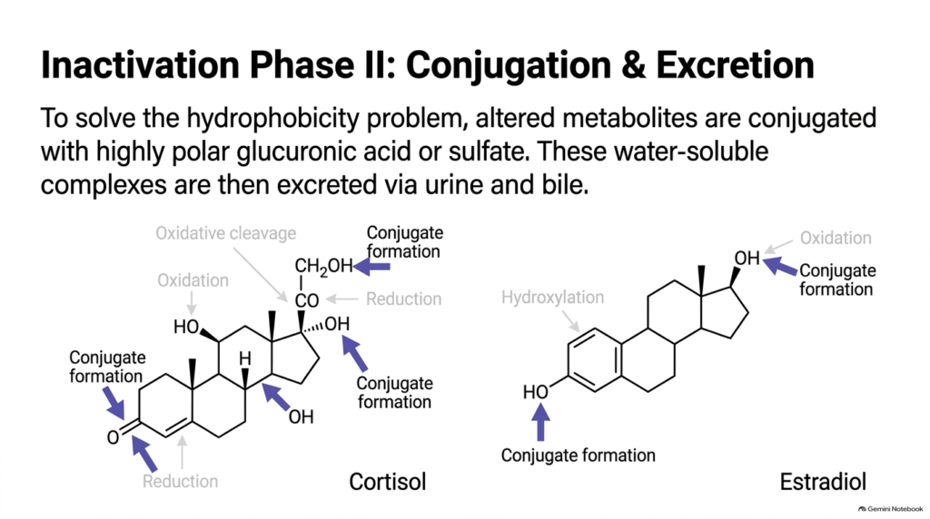
Following Phase I structural modifications, steroid hormone metabolism resolves the fundamental physical challenge of clearing hydrophobic molecules from the body. Even after losing biological activity, modified steroid metabolites remain relatively insoluble in aqueous blood plasma. Phase II inactivation in steroid hormone metabolism overcomes this obstacle by attaching highly polar chemical groups to modified steroids, successfully converting lipophilic metabolites into water-soluble compounds ready for rapid excretion through liquid waste pathways.
During Phase II conjugation, specialized liver enzymes transfer polar molecules onto hydroxyl groups created or exposed during Phase I. UDP-glucuronosyltransferases attach glucuronic acid moieties, while sulfotransferases attach sulfate groups. These conjugation reactions dramatically increase the water solubility of the steroid metabolites. Once conjugated, these polar complexes are efficiently transported out of hepatocytes into the bloodstream for urinary excretion by the kidneys or into bile for fecal elimination.
Mastering Phase II conjugation completes the study of steroid hormone metabolism. Excreted steroid conjugates measured in urine provide valuable clinical diagnostic biomarkers, allowing physicians to assess adrenal and gonadal hormone production non-invasively. By connecting Phase II excretion back to initial synthesis, students gain a complete end-to-end perspective on how the human body manages the entire lifecycle of steroid signaling molecules from origin to removal.
Slide 15: Steroid Hormone Metabolism – Summary Map of Structural Modifications
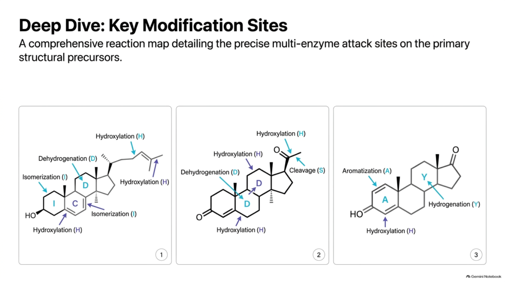
This final summary slide integrates all major concepts into a comprehensive reaction map of steroid hormone metabolism. By mapping the precise enzymatic attack sites across cholesterol, progesterone, and sex steroids, this visual overview consolidates the entire pathway into a single unified picture. It reinforces how specific functional-group modifications drive steroid hormone metabolism from precursor molecules to final active hormones and clearance products across different human organ systems.
Tracing reaction sites across the three primary precursor frames illustrates the elegant logic of steroid biochemistry. Hydroxylations at carbons 17, 21, and 11 define corticosteroid pathways, side-chain cleavages at carbons 20-22 and 17-20 control carbon loss, and A-ring modifications govern both progesterone activation and estrogen aromatization. Seeing these modification sites side by side allows students to compare how different enzyme families target distinct regions of the steroid nucleus during active hormone synthesis and metabolic regulation.
Reviewing this comprehensive reaction map provides the ultimate study tool for mastering steroid hormone metabolism. By synthesizing structural transformations, catalytic enzyme types, and organelle locations into one coherent framework, students transition from memorizing isolated reactions to understanding integrated endocrine physiology. This visual synthesis prepares students to excel on advanced biochemistry exams and confidently apply these core concepts in clinical practice and pathophysiology.
Please read our Content Disclaimer Statement.
Check out our social media channels: